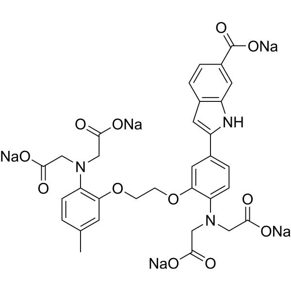
固绿 FCF
Fast Green FCF 是一种海绿色三芳基甲烷食用染料,最大吸收范围为 622 至 626 nm。Fast Green FCF 可抑制 α-synuclein 聚集,抑制 Aβ、P2X4 受体、TLR4/Myd88/NF-κB。Fast Green FCF 广泛用作染色剂,如在酸性提取 DNA 后在碱性 pH 下定量染色组蛋白,以及在电泳中用作蛋白质染色剂。Fast Green FCF 改善认知障碍、抑郁症、缓解疼痛过敏、促进生殖功能。
Fast green FCF free acid 是耐酸性的染料。Fast Green FCF free acid 可抑制 α-synuclein 聚集,抑制 Aβ、P2X4 受体、TLR4/Myd88/NF-κB。Fast Green FCF free acid 广泛用作染色剂,如在酸性提取 DNA 后在碱性 pH 下定量染色组蛋白,以及在电泳中用作蛋白质染色剂。Fast Green FCF free acid 改善认知障碍、抑郁症、缓解疼痛过敏、促进生殖功能。
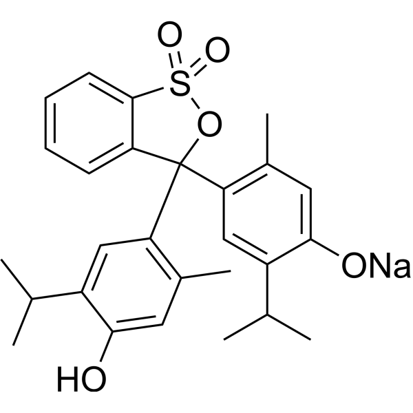
尿石素B (标准品)
Urolithin B (Standard) 是 Urolithin B 的分析标准品。本产品用于研究及分析应用。Urolithin B 是 Ellagitannins 的肠道微生物代谢产物之一,具有抗炎和抗氧化作用。Urolithin B 通过降低 IκBα 的磷酸化和降解来抑制 NF-κB 活性。Urolithin B 抑制 JNK、ERK 和 Akt 的磷酸化,增强 AMPK 的磷酸化。Urolithin B 也是骨骼肌质量的调节因子。
4-MUNANA (solution) 是一种流感病毒神经氨酸酶 (NA) 的底物,具有高选择性和反应不可逆性。在酶促反应中,4-MUNANA 被 NA 催化水解,生成具有荧光的 4-Methylumbelliferone (HY-N0187) ,通过检测 4-MU 的荧光强度,可实现对 NA 活性的定量分析。4-MUNANA 可用于流感相关研究,如筛选 NA 抑制剂,开发新型抗流感药物,研究流感病毒的感染机制。 溶剂及浓度:无菌水:10 mM
固绿 FCF
Fast Green FCF 是一种海绿色三芳基甲烷食用染料,最大吸收范围为 622 至 626 nm。Fast Green FCF 可抑制 α-synuclein 聚集,抑制 Aβ、P2X4 受体、TLR4/Myd88/NF-κB。Fast Green FCF 广泛用作染色剂,如在酸性提取 DNA 后在碱性 pH 下定量染色组蛋白,以及在电泳中用作蛋白质染色剂。Fast Green FCF 改善认知障碍、抑郁症、缓解疼痛过敏、促进生殖功能。
Fast green FCF free acid 是耐酸性的染料。Fast Green FCF free acid 可抑制 α-synuclein 聚集,抑制 Aβ、P2X4 受体、TLR4/Myd88/NF-κB。Fast Green FCF free acid 广泛用作染色剂,如在酸性提取 DNA 后在碱性 pH 下定量染色组蛋白,以及在电泳中用作蛋白质染色剂。Fast Green FCF free acid 改善认知障碍、抑郁症、缓解疼痛过敏、促进生殖功能。
4-MUNANA (solution) 是一种流感病毒神经氨酸酶 (NA) 的底物,具有高选择性和反应不可逆性。在酶促反应中,4-MUNANA 被 NA 催化水解,生成具有荧光的 4-Methylumbelliferone (HY-N0187) ,通过检测 4-MU 的荧光强度,可实现对 NA 活性的定量分析。4-MUNANA 可用于流感相关研究,如筛选 NA 抑制剂,开发新型抗流感药物,研究流感病毒的感染机制。 溶剂及浓度:无菌水:10 mM
尿石素B (标准品)
Urolithin B (Standard) 是 Urolithin B 的分析标准品。本产品用于研究及分析应用。Urolithin B 是 Ellagitannins 的肠道微生物代谢产物之一,具有抗炎和抗氧化作用。Urolithin B 通过降低 IκBα 的磷酸化和降解来抑制 NF-κB 活性。Urolithin B 抑制 JNK、ERK 和 Akt 的磷酸化,增强 AMPK 的磷酸化。Urolithin B 也是骨骼肌质量的调节因子。
Western blot analysis of extracts from THP-1(lane 2(20μg), Jurkat (lane 3(20μg) and NIH3T3(lane 4(20μg) using FOXO1A (HY-P80132) Rabbit mAb. Proteins were transferred
to a PVDF membrane and blocked with 5% non-fat milk in TBST for 2 hour at room temperature. The primary antibody (1/1000) and Loading control antibody (Beta Actin, HY-P80438, 1/10000) was
used in 5% non-fat milk in TBST at 4°C overnight. Goat Anti-Mouse/Rabbit IgG-HRP Secondary Antibody (1/10000) was used for 1 hour at room temperature.
Western blot analysis of extracts from THP-1(lane 2(20μg), Jurkat (lane 3(20μg) and NIH3T3(lane 4(20μg) using FOXO1A (HY-P80132) Rabbit mAb. Proteins were transferred
to a PVDF membrane and blocked with 5% non-fat milk in TBST for 2 hour at room temperature. The primary antibody (1/1000) and Loading control antibody (Beta Actin, HY-P80438, 1/10000) was
MedchemExpress Validation 03
Western blot analysis of extracts from THP-1(lane 2(20μg), Jurkat (lane 3(20μg) and NIH3T3(lane 4(20μg) using FOXO1A (HY-P80132) Rabbit mAb. Proteins were transferred
MedchemExpress Validation 04
Western blot analysis of extracts from THP-1(lane 2(20μg), Jurkat (lane 3(20μg) and NIH3T3(lane 4(20μg) using FOXO1A (HY-P80132) Rabbit mAb. Proteins were transferred
to a PVDF membrane and blocked with 5% non-fat milk in TBST for 2 hour at room temperature. The primary antibody (1/1000) and Loading control antibody (Beta Actin, HY-P80438, 1/10000) was
used in 5% non-fat milk in TBST at 4°C overnight. Goat Anti-Mouse/Rabbit IgG-HRP Secondary Antibody (1/10000) was used for 1 hour at room temperature.
MedchemExpress Validation
Western blot analysis of extracts from THP-1(lane 2(20μg), Jurkat (lane 3(20μg) and NIH3T3(lane 4(20μg) using FOXO1A (HY-P80132) Rabbit mAb. Proteins were transferred
to a PVDF membrane and blocked with 5% non-fat milk in TBST for 2 hour at room temperature. The primary antibody (1/1000) and Loading control antibody (Beta Actin, HY-P80438, 1/10000) was
used in 5% non-fat milk in TBST at 4°C overnight. Goat Anti-Mouse/Rabbit IgG-HRP Secondary Antibody (1/10000) was used for 1 hour at room temperature.
MedchemExpress Validation
Western blot analysis of extracts from THP-1(lane 2(20μg), Jurkat (lane 3(20μg) and NIH3T3(lane 4(20μg) using FOXO1A (HY-P80132) Rabbit mAb. Proteins were transferred
to a PVDF membrane and blocked with 5% non-fat milk in TBST for 2 hour at room temperature. The primary antibody (1/1000) and Loading control antibody (Beta Actin, HY-P80438, 1/10000) was
used in 5% non-fat milk in TBST at 4°C overnight. Goat Anti-Mouse/Rabbit IgG-HRP Secondary Antibody (1/10000) was used for 1 hour at room temperature.


![N-[3-(2-Furyl)acryloyl]-Phe-Gly-Gly](http://hg.y866.cn/biomolecule/lib/file/product_pic/hy-w010991.gif)