英文名称
CEA中文名称
癌胚抗原抗体别 名Carcinoembryonic antigen; Carcinoembryonic antigen related cell adhesion molecule 5; Carcinoembryonic antigen-related cell adhesion molecule 5; CD66e; CD66e; CD66e antigen; CEA; CEA; CEACAM5; CEAM5_HUMAN; DKFZp781M2392; Meconium antigen 100; OTTHUMP00000199032; OTTHUMP00000199033; OTTHUMP00000199034.
规格100ul 200ul
研究领域肿瘤 免疫学 干细胞 肿瘤细胞生物标志物
抗体来源Rabbit
克隆类型Polyclonal
交叉反应 Human, Rat,
产品应用WB=1:500-2000 ELISA=1:500-1000 IHC-P=1:400-800 IHC-F=1:400-800 Flow-Cyt=0.2μg/Test IF=1:100-500 (石蜡切片需做抗原修复)
not yet tested in other applications.
optimal dilutions/concentrations should be determined by the end user.
分 子 量71kDa
细胞定位细胞膜
性 状Lyophilized or Liquid
浓 度1mg/ml
免 疫 原KLH conjugated synthetic peptide derived from human CEA N-terminus:51-120/349
亚 型IgG
纯化方法affinity purified by Protein A
储 存 液0.01M TBS(pH7.4) with 1% BSA, 0.03% Proclin300 and 50% Glycerol.
保存条件Store at -20 °C for one year. Avoid repeated freeze/thaw cycles. The lyophilized antibody is stable at room temperature for at least one month and for greater than a year when kept at -20°C. When reconstituted in sterile pH 7.4 0.01M PBS or diluent of antibody the antibody is stable for at least two weeks at 2-4 °C.
PubMedPubMed
产品介绍background:
CEA-related cell adhesion molecules (CEACAM) belong to the carcinoembryonic antigen (CEA) family. It consists of seven CEACAM (CEACAM 1, CEACAM 3-CEACAM 8) and 11 pregnancy-specific glyco-protein (PSG 1-PSG 11) members. The CEA family proteins belong to the immunoglobulin (Ig) superfamily and are composed of one Ig variable-like (IgV) and a varying number (0-6) of Ig constant-like (IgC) domains. CEACAM molecules are membrane-bound either via a transmembrane domain or a glycosyl phosphatidyl inositol (GPI) anchor. CEACAM molecules are differentially expressed in epithelial cells or in leucocytes. Over-expression of CEA/ CEACAM 5 in tumors of epithelial origin is the basis of its wide-spread use as a tumor marker. The function of CEACAM family members varies widely: they function as cell adhesion molecules, tumor suppressors, regulators of lymphocyte and dendritic cell activation, receptors of Neisseria species and other bacteria.
Function:
Cell surface glycoprotein that plays a role in cell adhesion and in intracellular signaling. Receptor for E.coli Dr adhesins.
Subunit:
Homodimer. Binding of E.coli Dr adhesins leads to dissociation of the homodimer.
Subcellular Location:
Cell membrane; Lipid-anchor, GPI-anchor
Tissue Specificity:
Found in adenocarcinomas of endodermally derived digestive system epithelium and fetal colon.
Post-translational modifications:
Complex immunoreactive glycoprotein with a MW of 180 kDa comprising 60% carbohydrate.
Similarity:
Belongs to the immunoglobulin superfamily. CEA family.
Contains 7 Ig-like (immunoglobulin-like) domains.
SWISS:
P40199
Gene ID:
4680
Database links:
Entrez Gene: 4680 Human
Entrez Gene: 100125369 Rat
Omim: 163980 Human
SwissProt: P40199 Human
Unigene: 466814 Human
Unigene: 122781 Rat
Important Note:
This product as supplied is intended for research use only, not for use in human, therapeutic or diagnostic applications.
CEA是一种胚胎性抗原,主要存在于胎儿消化道上皮组织,胰腺和肝癌中。CEA广泛存在各种上皮性肿瘤,尤其是胃肠道恶性肿瘤,CEA分布阳性类型与肿瘤的恶性度有关。CEA与EMA一样可作为上皮性恶性肿瘤的重要标记。
| 产品图片 | 
Sample: SW480 Cell Lysate at 40 ug
Primary: Anti- CEA(bs-0060R) at 1/300 dilution
Secondary: IRDye800CW Goat Anti-Rabbit IgG at 1/10000 dilution
Predicted band size: 71 kD
Observed band size: 71 kD
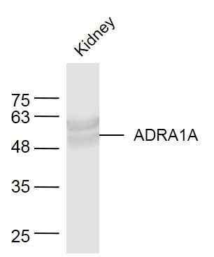
Sample:
Kidney (Mouse) Lysate at 40 ug
Primary: Anti-ADRA1A (bs-0060R) at 1/300 dilution
Secondary: IRDye800CW Goat Anti-Rabbit IgG at 1/20000 dilution
Predicted band size: 51 kD
Observed band size: 51 kD
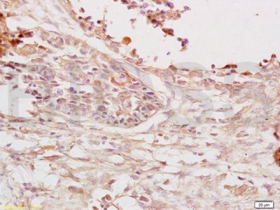
Tissue/cell: human lung carcinoma; 4% Paraformaldehyde-fixed and paraffin-embedded;
Antigen retrieval: citrate buffer ( 0.01M, pH 6.0 ), Boiling bathing for 15min; Block endogenous peroxidase by 3% Hydrogen peroxide for 30min; Blocking buffer (normal goat serum,C-0005) at 37℃ for 20 min;
Incubation: Anti-CEA/CD66e/CEACAM5 Polyclonal Antibody, Unconjugated(bs-0060R) 1:200, overnight at 4°C, followed by conjugation to the secondary antibody(SP-0023) and DAB(C-0010) staining
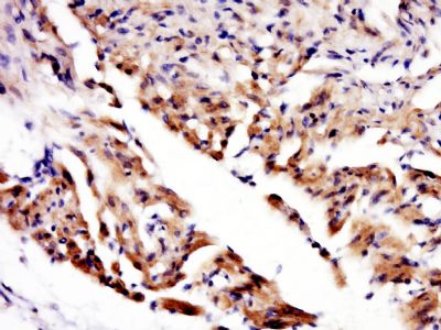
Paraformaldehyde-fixed, paraffin embedded (rat heart); Antigen retrieval by boiling in sodium citrate buffer (pH6.0) for 15min; Block endogenous peroxidase by 3% hydrogen peroxide for 20 minutes; Blocking buffer (normal goat serum) at 37°C for 30min; Antibody incubation with (ADRAIA) Polyclonal Antibody, Unconjugated (bs-0060R) at 1:400 overnight at 4°C, followed by a conjugated secondary (sp-0023) for 20 minutes and DAB staining.
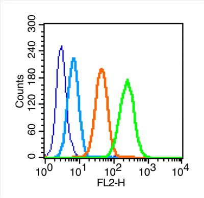
Blank control (blue line): MCF7 (blue).
Primary Antibody (green line): Rabbit Anti-CEA antibody (bs-0060R)
Dilution: 0.2μg /10^6 cells;
Isotype Control Antibody (orange line): Rabbit IgG .
Secondary Antibody (white blue line): Goat anti-rabbit IgG-PE
Dilution: 1μg /test.
Protocol
The cells were fixed with 70% methanol overnight at 4℃ . Cells stained with Primary Antibody for 30 min at room temperature. The cells were then incubated in 1 X PBS/2%BSA/10% goat serum to block non-specific protein-protein interactions followed by the antibody for 15 min at room temperature. The secondary antibody used for 40 min at room temperature. Acquisition of 20,000 events was performed. |


