| 亚型 | IgG |
| 形态 | 液态/冻干粉 |
| 保存条件 | -20℃保存 |
| 克隆性 | 多克隆 |
| 标记物 | FITC |
| 宿主 | Rabbit |
| 应用范围 | ELISA=1:500-1000 IHC-P=1:100-500 IHC-F=1:100-500 ICC=1:100-500 IF=1:100-2302 (石蜡切片需做抗原修复) not yet tested in other applications. |
| 浓度 | 1839mg/ml |
| 抗体英文名 | Cathepsin L |
| 规格 | 0.1ml 0.1804ml |
| 品图片 | 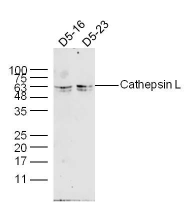 Sample: D5-16 Cell (Human) Lysate at 30 ug D5-23 Cell (Human) Lysate at 40 ug Primary: Anti- Cathepsin L (bs-1508R) at 1/300 dilution Secondary: IRDye800CW Goat Anti-Rabbit IgG at 1/20000 dilution Predicted band size: 19/30/37 kD Observed band size: 40/63 kD 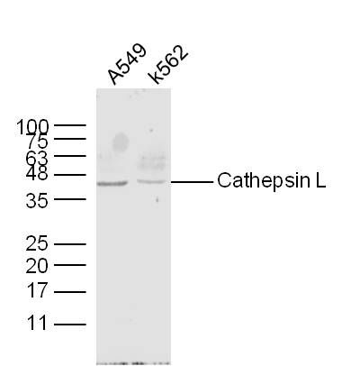 Sample: A549 Cell (Human) Lysate at 30 ug K562 Cell (Human) Lysate at 30 ug Primary: Anti- Cathepsin L (bs-1508R) at 1/300 dilution Secondary: IRDye800CW Goat Anti-Rabbit IgG at 1/20000 dilution Predicted band size: 19/30/37 kD Observed band size: 40/63 kD 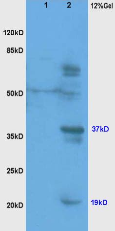 Protein: line1, rat brain lysates, 30ug; line2, rat liver lysates, 30ug; Primary: Anti-Cathepsin L (bs-1508R) at 1:200; Secondary: HRP conjugated Goat-Anti-Rabbit IgG(bs-0295G-HRP) at 1: 3000; ECL excitated the fluorescence; Predicted band size : 37kD Observed band size : 19kD, 37kD 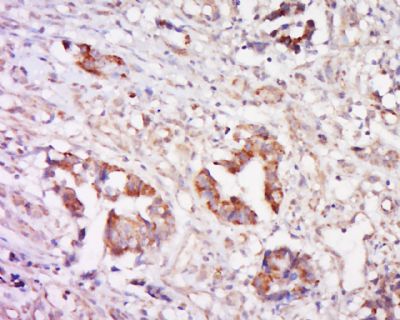 Tissue/cell: human cervical carcinoma; 4% Paraformaldehyde-fixed and paraffin-embedded; Antigen retrieval: citrate buffer ( 0.01M, pH 6.0 ), Boiling bathing for 15min; Block endogenous peroxidase by 3% Hydrogen peroxide for 30min; Blocking buffer (normal goat serum,C-0005) at 37℃ for 20 min; Incubation: Anti-Cathepsin Polyclonal Antibody, Unconjugated(bs-1508R) 1:500, overnight at 4°C, followed by conjugation to the secondary antibody(SP-0023) and DAB(C-0010) staining 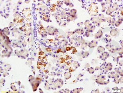 Tissue/cell: rat pancreas tissue; 4% Paraformaldehyde-fixed and paraffin-embedded; Antigen retrieval: citrate buffer ( 0.01M, pH 6.0 ), Boiling bathing for 15min; Block endogenous peroxidase by 3% Hydrogen peroxide for 30min; Blocking buffer (normal goat serum,C-0005) at 37℃ for 20 min; Incubation: Anti-Cathepsin L Polyclonal Antibody, Unconjugated(bs-1508R) 1:200, overnight at 4°C, followed by conjugation to the secondary antibody(SP-0023) and DAB(C-0010) staining |


