| 供应商 | 上海联迈生物工程有限公司 |
| 数量 | 大量 |
| 目录编号 | LM-1251R |
| 克隆性 | 多克隆 |
| 抗原来源 | Rabbit |
| 保质期 | 1年 |
| 抗体英文名 | CD166 |
| 抗体名 | 活化白细胞粘附分子抗体 |
| 宿主 | Rabbit |
| 适应物种 | Human, Mouse, Rat, Chicken, Dog, Pig, Cow, Horse, Rabbit, |
| 免疫原 | KLH conjugated synthetic peptide derived from human CD166:451-583/583 <Cytoplasmic> |
| 亚型 | IgG |
| 形态 | Lyophilized or Liquid |
| 应用范围 | WB=1:500-2000 ELISA=1:500-1000 IHC-F=1:400-800 Flow-Cyt=1μg/Test IF=1:100-500 (石蜡切片需做抗原修复) |
| 浓度 | 1mg/ml |
| 保存条件 | Store at -20 °C |
| 规格 | 50ul 100ul 200ul |
| 英文名称 | CD166 |
| 中文名称 | 活化白细胞粘附分子抗体 |
| 别 名 | activated leukocyte cell adhesion molecule; ALCAM protein; ALCAM; CD 166; CD166 antigen; FLJ38514; MEMD; MGC71733; FLJ3851; AI853494; BEN; DM-GRASP; MGC27910; MuSC; SC1; CD166_HUMAN. |
 | Specific References (2) | bs-1251R has been referenced in 2 publications. [IF=3.53] Lee, Tao-Chen, et al. "Comparison of Surface Markers between Human and Rabbit Mesenchymal Stem Cells." PLOS ONE 9.11 (2014): e111390. Rabbit. PubMed:25380245 [IF=1.99] Kovac, Michal, et al. "Different RNA and protein expression of surface markers in rabbit amniotic fluid‐derived mesenchymal stem cells." Biotechnology Progress (2017). FCM ; Rabbit. PubMed:28653478 |
| 规格价格 | 50ul/780元 购买 100ul/1380元 购买 200ul/2200元 购买 大包装/询价 |
| 说 明 书 | 50ul 100ul 200ul |
| 研究领域 | 肿瘤 细胞粘附分子 细胞表面分子 |
| 抗体来源 | Rabbit |
| 克隆类型 | Polyclonal |
| 交叉反应 | Human, Mouse, Rat, Chicken, Dog, Pig, Cow, Horse, Rabbit, |
| 产品应用 | WB=1:500-2000 ELISA=1:500-1000 IHC-F=1:400-800 Flow-Cyt=1μg/Test IF=1:100-500 (石蜡切片需做抗原修复) not yet tested in other applications. optimal dilutions/concentrations should be determined by the end user. |
| 分 子 量 | 62kDa |
| 细胞定位 | 细胞膜 |
| 性 状 | Lyophilized or Liquid |
| 浓 度 | 1mg/ml |
| 免 疫 原 | KLH conjugated synthetic peptide derived from human CD166:451-583/583 <Cytoplasmic> |
| 亚 型 | IgG |
| 纯化方法 | affinity purified by Protein A |
| 储 存 液 | 0.01M TBS(pH7.4) with 1% BSA, 0.03% Proclin300 and 50% Glycerol. |
| 保存条件 | Store at -20 °C for one year. Avoid repeated freeze/thaw cycles. The lyophilized antibody is stable at room temperature for at least one month and for greater than a year when kept at -20°C. When reconstituted in sterile pH 7.4 0.01M PBS or diluent of antibody the antibody is stable for at least two weeks at 2-4 °C. |
| PubMed | PubMed |
| 产品介绍 | background: CD166 is a member of the Ig superfamily and is expressed on activated T-cells, B cells and other cells including thymic epithelial cells, fibroblasts, keratinocytes and neurons. CD6 has been identified as a receptor for CD166. The expression of CD166 is up-regulated in low-grade prostate tumors and down-regulated in high-grade tumors; may play role in progression of prostate cancer. Function: Cell adhesion molecule that binds to CD6. Involved in neurite extension by neurons via heterophilic and hemophilic interactions. May play a role in the binding of T- and B-cells to activated leukocytes, as well as in interactions between cells of the nervous system. Subcellular Location: Membrane; Single-pass type I membrane protein. Tissue Specificity: Spleen, placenta, liver, and weakly in liver. Expressed by activated T-cells, B-cells, monocytes and thymic epithelial cells. Expressed by neurons in the brain. Restricted expression in tumor cell lines. Preferentially expressed in highly metastasizing melanoma cell lines. Similarity: Contains 3 Ig-like C2-type (immunoglobulin-like) domains. Contains 2 Ig-like V-type (immunoglobulin-like) domains. SWISS: Q13740 Gene ID: 214 Database links: Entrez Gene: 214 Human Entrez Gene: 11658 Mouse Entrez Gene: 79559 Rat Omim: 601662 Human SwissProt: Q13740 Human SwissProt: Q61490 Mouse SwissProt: O35112 Rat Unigene: 591293 Human Unigene: 288282 Mouse Unigene: 5789 Rat Important Note: This product as supplied is intended for research use only, not for use in human, therapeutic or diagnostic applications. ALCAM/CD166是免疫球蛋白超家族的成员,对多种肿瘤生长和转移相关的肿瘤细胞特性具有调节作用,ALCAM/CD166在乳腺癌、前列腺癌、食道癌、结肠癌及恶性黑色素瘤等肿瘤都有一定的表达,尤以结肠、直肠癌表达显著。 |
| 产品图片 | 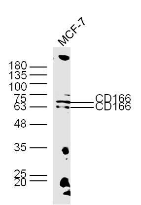 Sample: MCF-7 Cell (Human) Lysate at 30 ug Primary: Anti-CD166 (bs-1251R) at 1/300 dilution Secondary: IRDye800CW Goat Anti-Rabbit IgG at 1/20000 dilution Predicted band size: 62 kD Observed band size: 62/70 kD 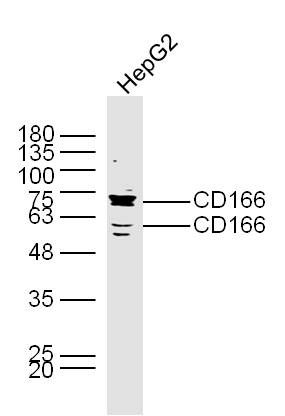 Sample: HepG2 Cell (Human) Lysate at 30 ug Primary: Anti-CD166 (bs-1251R) at 1/300 dilution Secondary: IRDye800CW Goat Anti-Rabbit IgG at 1/20000 dilution Predicted band size: 62 kD Observed band size: 62/70 kD 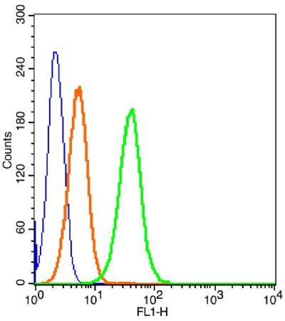 Blank control: Molt-4 Cells(blue). Primary Antibody: Rabbit Anti-CD166/FITC Conjugated antibody (bs-1251R/AF488), Dilution: 1μg in 100 μL 1X PBS containing 0.5% BSA; Isotype Control Antibody: Rabbit IgG/AF488orange) ,used under the same conditions. Protocol The cells were fixed with 2% paraformaldehyde (10 min) . The cells were washed twice with 1 X PBS. The cells were incubated in 1 X PBS containing 0.5% BSA + 1 0% goat serum (15 min) to block non-specific protein-protein interactions followed by the incubated with antibody (bs-1251R/AF488, 1μg /1x10^6 cells) for 30 min on ice. Acquisition of 20,000 events was performed. 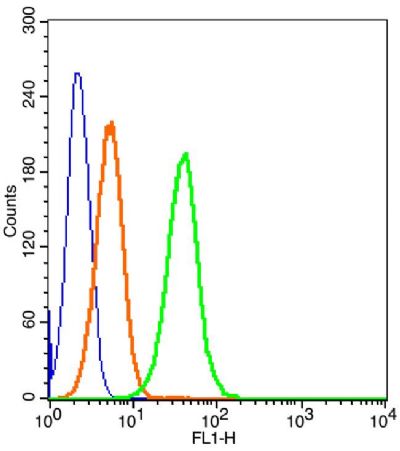 Blank control: Molt-4 Cells(blue). Primary Antibody: Rabbit Anti-CD166/FITC Conjugated antibody (bs-1251R/AF488), Dilution: 1μg in 100 μL 1X PBS containing 0.5% BSA; Isotype Control Antibody: Rabbit IgG/AF488orange) ,used under the same conditions. Protocol The cells were fixed with 2% paraformaldehyde (10 min) . The cells were washed twice with 1 X PBS. The cells were incubated in 1 X PBS containing 0.5% BSA + 1 0% goat serum (15 min) to block non-specific protein-protein interactions followed by the incubated with antibody (bs-1251R/AF488, 1μg /1x10^6 cells) for 30 min on ice. Acquisition of 20,000 events was performed. |


