| 供应商 | 上海联迈生物工程有限公司 |
| 数量 | 大量 |
| 目录编号 | LM-1424R |
| 克隆性 | 多克隆 |
| 抗原来源 | Rabbit |
| 保质期 | 1年 |
| 抗体英文名 | EPOR |
| 抗体名 | 红细胞生成素受体抗体 |
| 宿主 | Rabbit |
| 适应物种 | Human, Mouse, Rat, Dog, Cow, Horse, |
| 免疫原 | KLH conjugated synthetic peptide derived from human EPOR:301-450/508 <Cytoplasmic> |
| 亚型 | IgG |
| 形态 | Lyophilized or Liquid |
| 应用范围 | WB=1:500-2000 ELISA=1:500-1000 IHC-P=1:400-800 IHC-F=1:400-800 Flow-Cyt=1µg/Test IF=1:100-500 (石蜡切片需做抗原修复) |
| 浓度 | 1mg/ml |
| 保存条件 | Store at -20 °C |
| 规格 | 50ul 100ul 200ul |
| 英文名称 | EPOR |
| 中文名称 | 红细胞生成素受体抗体 |
| 别 名 | erythropoietin receptor; EPO R; EPO Receptor; Erythropoietin receptor precursor; EPOR_HUMAN; MGC138358. |
| 规格价格 | 50ul/780元 购买 100ul/1380元 购买 200ul/2200元 购买 大包装/询价 |
| 说 明 书 | 50ul 100ul 200ul |
| 研究领域 | 肿瘤 心血管 免疫学 信号转导 干细胞 细胞凋亡 细胞膜受体 G蛋白信号 |
| 抗体来源 | Rabbit |
| 克隆类型 | Polyclonal |
| 交叉反应 | Human, Mouse, Rat, Dog, Cow, Horse, |
| 产品应用 | WB=1:500-2000 ELISA=1:500-1000 IHC-P=1:400-800 IHC-F=1:400-800 Flow-Cyt=1µg/Test IF=1:100-500 (石蜡切片需做抗原修复) not yet tested in other applications. optimal dilutions/concentrations should be determined by the end user. |
| 分 子 量 | 56kDa |
| 细胞定位 | 细胞膜 细胞外基质 分泌型蛋白 |
| 性 状 | Lyophilized or Liquid |
| 浓 度 | 1mg/ml |
| 免 疫 原 | KLH conjugated synthetic peptide derived from human EPOR:301-450/508 <Cytoplasmic> |
| 亚 型 | IgG |
| 纯化方法 | affinity purified by Protein A |
| 储 存 液 | 0.01M TBS(pH7.4) with 1% BSA, 0.03% Proclin300 and 50% Glycerol. |
| 保存条件 | Store at -20 °C for one year. Avoid repeated freeze/thaw cycles. The lyophilized antibody is stable at room temperature for at least one month and for greater than a year when kept at -20°C. When reconstituted in sterile pH 7.4 0.01M PBS or diluent of antibody the antibody is stable for at least two weeks at 2-4 °C. |
| PubMed | PubMed |
| 产品介绍 | background: The erythropoietin receptor (EPOR) is a member of the cytokine receptor family. There are several isoforms including: EPOR-F (full length), EPOR-S (soluble form), and EPOR-T (truncated form). Upon erythropoietin (EPO) binding, the EPOR activates Jak2 tyrosine kinase which activates different intracellular pathways including: Ras/MAP kinase, phosphatidylinositol 3-kinase and STAT transcription factors. The stimulated EPOR appears to have a role in erythroid cell survival. Defects in the EPOR may produce erythroleukemia and familial erythrocytosis. A functional EPOR is found in the cardiovascular system, including endothelial cells and cardiomyocytes, and data suggest that the EPO/EPO receptor system plays an important role in cardiac function. In animal studies, treatment with EPO during ischemia/reperfusion in the heart has been shown to limit the infarct size and the extent of apoptosis. Function: Receptor for erythropoietin. Mediates erythropoietin-induced erythroblast proliferation and differentiation. Upon EPO stimulation, EPOR dimerizes triggering the JAK2/STAT5 signaling cascade. In some cell types, can also activate STAT1 and STAT3. May also activate the LYN tyrosine kinase. Isoform EPOR-T acts as a dominant-negative receptor of EPOR-mediated signaling. Subunit: Forms homodimers on EPO stimulation. The tyrosine-phosphorylated form interacts with several SH2 domain-containing proteins including LYN, the adapter protein APS, PTPN6, PTPN11, JAK2, PI3 kinases, STAT5A/B, SOCS3, CRKL. Interacts with INPP5D/SHIP1. The N-terminal SH2 domain of PTPN6 binds Tyr-454 and inhibits signaling through dephosphorylation of JAK2. APS binding also inhibits the JAK-STAT signaling. Binding to PTPN11, preferentially through the N-terminal SH2 domain, promotes mitogenesis and phosphorylation of PTPN11. Binding of JAK2 (through its N-terminal) promotes cell-surface expression. Interaction with the ubiquitin ligase NOSIP mediates EPO-induced cell proliferation. Interacts with ATXN2L. Subcellular Location: Cell membrane; Single-pass type I membrane protein. Isoform EPOR-S: Secreted. Note=Secreted and located to the cell surface. Tissue Specificity: Erythroid cells and erythroid progenitor cells. Isoform EPOR-F is the most abundant form in EPO-dependent erythroleukemia cells and in late-stage erythroid progenitors. Isoform EPOR-S and isoform EPOR-T are the predominant forms in bone marrow. Isoform EPOR-T is the most abundant from in early-stage erythroid progenitor cells. Similarity: Belongs to the type I cytokine receptor family. Type 1 subfamily. Contains 1 fibronectin type-III domain. SWISS: P19235 Gene ID: 2057 Database links: Entrez Gene: 2057 Human Entrez Gene: 13857 Mouse Entrez Gene: 24336 Rat Omim: 133171 Human SwissProt: P19235 Human SwissProt: P14753 Mouse SwissProt: Q07303 Rat Unigene: 631624 Human Unigene: 2653 Mouse Unigene: 22394 Rat Important Note: This product as supplied is intended for research use only, not for use in human, therapeutic or diagnostic applications. |
| 产品图片 | 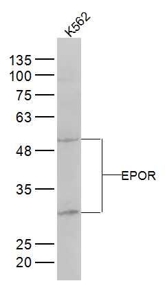 Sample: K562(Human) Cell Lysate at 30 ug Primary: Anti-EPOR (bs-1424R) at 1/300 dilution Secondary: IRDye800CW Goat Anti-Rabbit IgG at 1/20000 dilution Predicted band size: 56 kD Observed band size: 30/52 kD 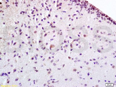 Tissue/cell: rat brain tissue; 4% Paraformaldehyde-fixed and paraffin-embedded; Antigen retrieval: citrate buffer ( 0.01M, pH 6.0 ), Boiling bathing for 15min; Block endogenous peroxidase by 3% Hydrogen peroxide for 30min; Blocking buffer (normal goat serum,C-0005) at 37℃ for 20 min; Incubation: Anti-EPOR Polyclonal Antibody, Unconjugated(bs-1424R) 1:100, overnight at 4°C, followed by conjugation to the secondary antibody(SP-0023) and DAB(C-0010) staining 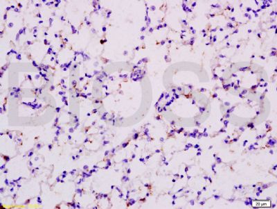 Tissue/cell: rat lung tissue; 4% Paraformaldehyde-fixed and paraffin-embedded; Antigen retrieval: citrate buffer ( 0.01M, pH 6.0 ), Boiling bathing for 15min; Block endogenous peroxidase by 3% Hydrogen peroxide for 30min; Blocking buffer (normal goat serum,C-0005) at 37℃ for 20 min; Incubation: Anti-EPOR Polyclonal Antibody, Unconjugated(bs-1424R) 1:200, overnight at 4°C, followed by conjugation to the secondary antibody(SP-0023) and DAB(C-0010) staining 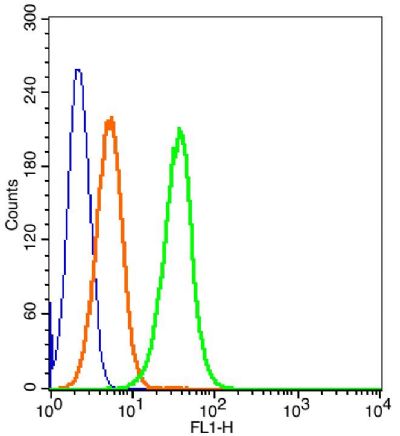 Blank control: Molt-4 Cells(blue). Primary Antibody: Rabbit Anti-EPOR/FITC Conjugated antibody (bs-1424R/AF488), Dilution: 1μg in 100 μL 1X PBS containing 0.5% BSA; Isotype Control Antibody: Rabbit IgG/AF488 orange) ,used under the same conditions. Protocol The cells were fixed with 2% paraformaldehyde (10 min) . The cells were washed twice with 1 X PBS. The cells were incubated in 1 X PBS containing 0.5% BSA + 1 0% goat serum (15 min) to block non-specific protein-protein interactions followed by the incubated with antibody (bs-1424R/AF488, 1μg /1x10^6 cells) for 30 min on ice. Acquisition of 20,000 events was performed. |


