找化学品上960化工网!
\n

Sandwich ELISA detection of non-transfected and transfected 293T whole cell extracts using GTX112677 as capture antibody at concentration of 5 μg/mL and a mouse monoclonal anti-APP antibody as detection antibody at concentration of 1 μg/mL. Mouse IgG antibody (HRP) (GTX213111-01) was diluted at 1:10000 and used to detect the primary antibody.

Rat tissue extract (50 μg) was separated by 7.5% SDS-PAGE, and the membrane was blotted with APP antibody (GTX112677) diluted at 1:500.

APP antibody detects APP protein at cytoplasm by immunofluorescent analysis.
Sample: SH-SY5Y cells were fixed in 4% paraformaldehyde at RT for 15 min.
Green: APP protein stained by APP antibody (GTX112677) diluted at 1:100.
Red: beta Tubulin 3/ TUJ1 protein stained by beta Tubulin 3/ TUJ1 antibody (GTX631836) diluted at 1:200.
Blue: Hoechst 33342 staining.

Various whole cell extracts (30 μg) were separated by 7.5% SDS-PAGE, and the membrane was blotted with APP antibody (GTX112677) diluted at 1:500.
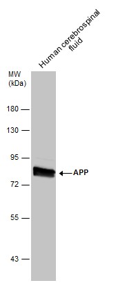
Human tissue extract (30 μg) was separated by 7.5% SDS-PAGE, and the membrane was blotted with APP antibody (GTX112677) diluted at 1:500.

APP antibody detects APP protein at cytoplasm by immunofluorescent analysis.
Sample: U-87 MG cells were fixed in 4% paraformaldehyde at RT for 15 min.
Green: APP protein stained by APP antibody (GTX112677) diluted at 1:100.
Blue: Hoechst 33342 staining.

APP antibody detects APP protein at cytoplasm in Rat brain by immunohistochemical analysis.
Sample: Paraffin-embedded Rat brain.
APP antibody (GTX112677) diluted at 1:500.
Antigen Retrieval: Citrate buffer, pH 6.0, 15 min

Mouse tissue extract (50 μg) was separated by 7.5% SDS-PAGE, and the membrane was blotted with APP antibody (GTX112677) diluted at 1:500.
Sandwich ELISA detection of non-transfected and transfected 293T whole cell extracts using GTX112677 as capture antibody at concentration of 5 μg/mL and a mouse monoclonal anti-APP antibody as detection antibody at concentration of 1 μg/mL. Mouse IgG antibody (HRP) (GTX213111-01) was diluted at 1:10000 and used to detect the primary antibody.
Sandwich ELISA detection of non-transfected and transfected 293T whole cell extracts using GTX112677 as capture antibody at concentration of 5 μg/mL and a mouse monoclonal anti-APP antibody as detection antibody at concentration of 1 μg/mL. Mouse IgG antibody (HRP) (GTX213111-01) was diluted at 1:10000 and used to detect the primary antibody.
Sandwich ELISA detection of non-transfected and transfected 293T whole cell extracts using GTX112677 as capture antibody at concentration of 5 μg/mL and a mouse monoclonal anti-APP antibody as detection antibody at concentration of 1 μg/mL. Mouse IgG antibody (HRP) (GTX213111-01) was diluted at 1:10000 and used to detect the primary antibody.Rat tissue extract (50 μg) was separated by 7.5% SDS-PAGE, and the membrane was blotted with APP antibody (GTX112677) diluted at 1:500.
Rat tissue extract (50 μg) was separated by 7.5% SDS-PAGE, and the membrane was blotted with APP antibody (GTX112677) diluted at 1:500.
Rat tissue extract (50 μg) was separated by 7.5% SDS-PAGE, and the membrane was blotted with APP antibody (GTX112677) diluted at 1:500.APP antibody detects APP protein at cytoplasm by immunofluorescent analysis.
Sample: SH-SY5Y cells were fixed in 4% paraformaldehyde at RT for 15 min.
Green: APP protein stained by APP antibody (GTX112677) diluted at 1:100.
Red: beta Tubulin 3/ TUJ1 protein stained by beta Tubulin 3/ TUJ1 antibody (GTX631836) diluted at 1:200.
Blue: Hoechst 33342 staining.
APP antibody detects APP protein at cytoplasm by immunofluorescent analysis.
Sample: SH-SY5Y cells were fixed in 4% paraformaldehyde at RT for 15 min.
Green: APP protein stained by APP antibody (GTX112677) diluted at 1:100.
Red: beta Tubulin 3/ TUJ1 protein stained by beta Tubulin 3/ TUJ1 antibody (GTX631836) diluted at 1:200.
Blue: Hoechst 33342 staining.
Various whole cell extracts (30 μg) were separated by 7.5% SDS-PAGE, and the membrane was blotted with APP antibody (GTX112677) diluted at 1:500.
Various whole cell extracts (30 μg) were separated by 7.5% SDS-PAGE, and the membrane was blotted with APP antibody (GTX112677) diluted at 1:500.
Various whole cell extracts (30 μg) were separated by 7.5% SDS-PAGE, and the membrane was blotted with APP antibody (GTX112677) diluted at 1:500.Human tissue extract (30 μg) was separated by 7.5% SDS-PAGE, and the membrane was blotted with APP antibody (GTX112677) diluted at 1:500.
Human tissue extract (30 μg) was separated by 7.5% SDS-PAGE, and the membrane was blotted with APP antibody (GTX112677) diluted at 1:500.
Human tissue extract (30 μg) was separated by 7.5% SDS-PAGE, and the membrane was blotted with APP antibody (GTX112677) diluted at 1:500.APP antibody detects APP protein at cytoplasm by immunofluorescent analysis.
Sample: U-87 MG cells were fixed in 4% paraformaldehyde at RT for 15 min.
Green: APP protein stained by APP antibody (GTX112677) diluted at 1:100.
Blue: Hoechst 33342 staining.
APP antibody detects APP protein at cytoplasm by immunofluorescent analysis.
Sample: U-87 MG cells were fixed in 4% paraformaldehyde at RT for 15 min.
Green: APP protein stained by APP antibody (GTX112677) diluted at 1:100.
Blue: Hoechst 33342 staining.
APP antibody detects APP protein at cytoplasm in Rat brain by immunohistochemical analysis.
Sample: Paraffin-embedded Rat brain.
APP antibody (GTX112677) diluted at 1:500.
Antigen Retrieval: Citrate buffer, pH 6.0, 15 min
APP antibody detects APP protein at cytoplasm in Rat brain by immunohistochemical analysis.
Sample: Paraffin-embedded Rat brain.
APP antibody (GTX112677) diluted at 1:500.
Antigen Retrieval: Citrate buffer, pH 6.0, 15 min
Mouse tissue extract (50 μg) was separated by 7.5% SDS-PAGE, and the membrane was blotted with APP antibody (GTX112677) diluted at 1:500.
Mouse tissue extract (50 μg) was separated by 7.5% SDS-PAGE, and the membrane was blotted with APP antibody (GTX112677) diluted at 1:500.
Mouse tissue extract (50 μg) was separated by 7.5% SDS-PAGE, and the membrane was blotted with APP antibody (GTX112677) diluted at 1:500.


