英文名称PCNA
中文名称
增殖细胞核抗原抗体别 名Proliferation Marker; Cyclin; DNA polymerase delta auxiliary protein; HGCN8729; MGC8367; Mutagen-sensitive 209 protein; Pcna/cyclin; PCNAR; Polymerase delta accessory protein; Proliferating Cell Nuclear Antigen; PCNA_HUMAN.
规格价格50ul/780元 购买 100ul/1380元 购买 200ul/2200元 购买 大包装/询价
说 明 书50ul 100ul 200ul
研究领域肿瘤 细胞生物 免疫学 染色质和核信号 细胞周期蛋白 细胞分化 细胞类型标志物
抗体来源Rabbit
克隆类型Polyclonal
交叉反应 Human, Mouse, Rat, Chicken, Dog, Cow, Rabbit,
产品应用WB=1:500-2000 ELISA=1:500-1000 IHC-P=1:400-800 IHC-F=1:400-800 Flow-Cyt=1μg/Test IF=1:100-500 (石蜡切片需做抗原修复)
not yet tested in other applications.
optimal dilutions/concentrations should be determined by the end user.
分 子 量29kDa
性 状Lyophilized or Liquid
浓 度1mg/ml
免 疫 原KLH conjugated synthetic peptide derived from human PCNA:201-261/261
亚 型IgG
纯化方法affinity purified by Protein A
储 存 液0.01M TBS(pH7.4) with 1% BSA, 0.03% Proclin300 and 50% Glycerol.
保存条件Store at -20 °C for one year. Avoid repeated freeze/thaw cycles. The lyophilized antibody is stable at room temperature for at least one month and for greater than a year when kept at -20°C. When reconstituted in sterile pH 7.4 0.01M PBS or diluent of antibody the antibody is stable for at least two weeks at 2-4 °C.
PubMedPubMed
产品介绍background:
Proliferation Marker
Proliferating cell nuclear antigen (PCNA) is a 28kDa nuclear protein associated with the cell cycle, a nuclear protein vital for cellular DNA synthesis. Proliferating cell nuclear antigen was originally identified by immunofluorescence as a nuclear protein whose appearance correlated with the proliferate state of the cell. PCNA is required for replication of DNA in vitro and has been identified as the auxiliary protein (cofactor) for DNA polymerase delta. The anti-PCNA antibodies react with the nuclei of proliferating cells. PCNA is essential for cellular DNA synthesis and is also required for the in vitro replication of simian virus 40 (SV40) DNA where it acts to coordinate leading and lagging strand synthesis at the replication fork. The PCNA protein may fulfil several separate roles in the cell nucleus associated with changes in its antigenic structure。
Function:
Auxiliary protein of DNA polymerase delta and is involved in the control of eukaryotic DNA replication by increasing the polymerase's processibility during elongation of the leading strand. Induces a robust stimulatory effect on the 3'-5' exonuclease and 3'-phosphodiesterase, but not apurinic-apyrimidinic (AP) endonuclease, APEX2 activities. Has to be loaded onto DNA in order to be able to stimulate APEX2. Plays a key role in DNA damage response (DDR) by being conveniently positioned at the replication fork to coordinate DNA replication with DNA repair and DNA damage tolerance pathways. Acts as a loading platform to recruit DDR proteins that allow completion of DNA replication after DNA damage and promote postreplication repair: Monoubiquitinated PCNA leads to recruitment of translesion (TLS) polymerases, while 'Lys-63'-linked polyubiquitination of PCNA is involved in error-free pathway and employs recombination mechanisms to synthesize across the lesion.
Subunit:
Homotrimer (By similarity). Forms a complex with activator 1 heteropentamer in the presence of ATP. Interacts with EXO1, POLH, POLK, DNMT1, ERCC5, FEN1, CDC6 and POLDIP2. Interacts with APEX2; this interaction is triggered by reactive oxygen species and increased by misincorporation of uracil in nuclear DNA. Forms a ternary complex with DNTTIP2 and core histone. Interacts with KCTD10 and PPP1R15A (By similarity). Interacts with POLD1, POLD3 and POLD4. Interacts with BAZ1B; the interaction is direct. Interacts with HLTF and SHPRH. Interacts with NUDT15. Interaction is disrupted in response to UV irradiation and acetylation. Interacts with CDKN1A/p21(CIP1) and CDT1; interacts via their PIP-box which also recruits the DCX(DTL) complex. Interacts with DDX11. Interacts with EGFR; positively regulates PCNA. Interacts with PARPBP. Interacts (when ubiquitinated) with SPRTN; leading to enhance RAD18-mediated PCNA ubiquitination. Interacts (when polyubiquitinated) with ZRANB3. Interacts with SMARCAD1. Interacts with CDKN1C. Interacts with KIAA0101/PAF15 (via PIP-box).
Subcellular Location:
Nucleus. Note=Forms nuclear foci representing sites of ongoing DNA replication and vary in morphology and number during S phase. Together with APEX2, is redistributed in discrete nuclear foci in presence of oxidative DNA damaging agents.
Post-translational modifications:
Following DNA damage, can be either monoubiquitinated to stimulate direct bypass of DNA lesions by specialized DNA polymerases or polyubiquitinated to promote recombination-dependent DNA synthesis across DNA lesions by template switching mechanisms. Following induction of replication stress, monoubiquitinated by the UBE2B-RAD18 complex on Lys-164, leading to recruit translesion (TLS) polymerases, which are able to synthesize across DNA lesions in a potentially error-prone manner. An error-free pathway also exists and requires non-canonical polyubiquitination on Lys-164 through 'Lys-63' linkage of ubiquitin moieties by the E2 complex UBE2N-UBE2V2 and the E3 ligases, HLTF, RNF8 and SHPRH. This error-free pathway, also known as template switching, employs recombination mechanisms to synthesize across the lesion, using as a template the undamaged, newly synthesized strand of the sister chromatid. Monoubiquitination at Lys-164 also takes place in undamaged proliferating cells, and is mediated by the DCX(DTL) complex, leading to enhance PCNA-dependent translesion DNA synthesis. Sumoylated during S phase.
Acetylated in response to UV irradiation. Acetylation disrupts interaction with NUDT15 and promotes degradation.
Phosphorylated. Phosphorylation at Tyr-211 by EGFR stabilizes chromatin-associated PCNA.
Similarity:
Belongs to the PCNA family.
SWISS:
P12004
Gene ID:
5111
Database links:
Entrez Gene: 515499 Cow
Entrez Gene: 5111 Human
Entrez Gene: 18538 Mouse
Entrez Gene: 25737 Rat
Omim: 176740 Human
SwissProt: Q3ZBW4 Cow
SwissProt: P12004 Human
SwissProt: P17918 Mouse
SwissProt: P04961 Rat
Unigene: 147433 Human
Unigene: 728886 Human
Unigene: 7141 Mouse
Unigene: 223 Rat
Important Note:
This product as supplied is intended for research use only, not for use in human, therapeutic or diagnostic applications.
PCNA是一种仅在增殖细胞中合成或表达的核内多肽,其表达和合成与细胞周期有关。主要表达于增殖细胞的S期、G1期和G2初期。
PCNA主要作为判断各种恶性肿瘤(包括胃肠道癌肿、乳腺癌、肝癌、膀胱癌等)细胞增殖和其恶性程度的一种指标.
Sample:
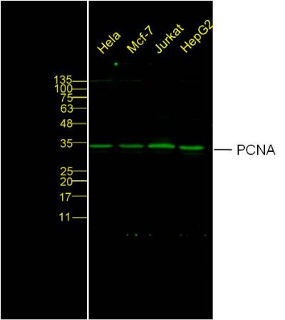
Hela Cell Lysate at 40 ug
MCF-7 Cell Lysate at 40 ug
Jurkat Cell Lysate at 40 ug
HepG2 Cell Lysate at 40 ug
Primary: Anti- PCNA(bs-2007R) at 1/2000 dilution
Secondary: IRDye800CW Goat Anti-Rabbit IgG at 1/20000 dilution
Predicted band size: 29 kD
Observed band size: 34 kD
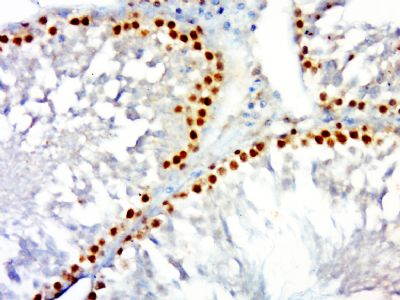
Paraformaldehyde-fixed, paraffin embedded (rat testis); Antigen retrieval by boiling in sodium citrate buffer (pH6.0) for 15min; Block endogenous peroxidase by 3% hydrogen peroxide for 20 minutes; Blocking buffer (normal goat serum) at 37°C for 30min; Antibody incubation with (PCNA) Polyclonal Antibody, Unconjugated (bs-2007R) at 1:500 overnight at 4°C, followed by a conjugated secondary (sp-0023) for 20 minutes and DAB staining.
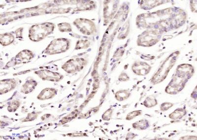
Paraformaldehyde-fixed, paraffin embedded (human gastric cancer); Antigen retrieval by boiling in sodium citrate buffer (pH6.0) for 15min; Block endogenous peroxidase by 3% hydrogen peroxide for 20 minutes; Blocking buffer (normal goat serum) at 37°C for 30min; Antibody incubation with (PCNA) Polyclonal Antibody, Unconjugated (bs-2007R) at 1:400 overnight at 4°C, followed by a conjugated secondary (sp-0023) for 20 minutes and DAB staining.
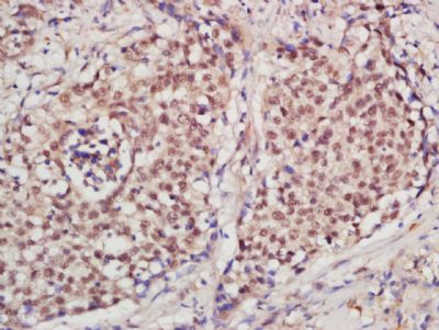
Tissue/cell: human laryngo carcinoma; 4% Paraformaldehyde-fixed and paraffin-embedded;
Antigen retrieval: citrate buffer ( 0.01M, pH 6.0 ), Boiling bathing for 15min; Block endogenous peroxidase by 3% Hydrogen peroxide for 30min; Blocking buffer (normal goat serum,C-0005) at 37℃ for 20 min;
Incubation: Anti-PCNA Polyclonal Antibody, Unconjugated(bs-2007R) 1:200, overnight at 4°C, followed by conjugation to the secondary antibody(SP-0023) and DAB(C-0010) staining
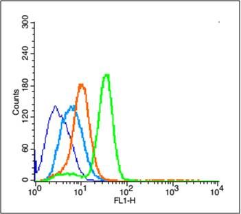
sample: A549 cells( (blue) Ice-cold 70% ethanol was added and the cells were incubated for overnight at 4 °C. and then resuspended in ice-cold 90% methanol for 30 min on ice.) Primary Antibody:Rabbit Anti-Phospho-NFKB1 (Ser373) antibody(bs-20075R) (green) Isotype Control Antibody: Rabbit IgG (orange) Secondary Antibody: F(ab’)2 fragment goat anti-rabbit IgG-FITC (white blue)
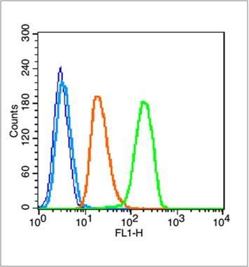
Blank control (blue line): U251 (blue).
Primary Antibody (green line): Rabbit Anti-PCNA antibody (bs-2007R)
Dilution: 3μg /10^6 cells;
Isotype Control Antibody (orange line): Rabbit IgG .
Secondary Antibody (white blue line): Goat anti-rabbit IgG-PE
Dilution: 1μg /test.
Protocol
The cells were fixed with 2% paraformaldehyde (10 min)and then permeabilized with 0.1% PBS-Tween for 20 min at room temperature. Cells stained with Primary Antibody for 30 min at room temperature. The cells were then incubated in 1 X PBS/2%BSA/10% goat serum to block non-specific protein-protein interactions followed by the antibody for 15 min at room temperature. The secondary antibody used for 40 min at room temperature. Acquisition of 20,000 events was performed.


