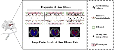
The development of an effective method for staging liver fibrosis has always been a hot topic of research in the field of liver fibrosis. In this paper, PEGylated ultrafine superparamagnetic iron oxide nanocrystals (SPIO@PEG) were developed for T1–T2 dual-modal contrast-enhanced magnetic resonance imaging (MRI) and combined with Matrix Laboratory (MATLAB)-based image fusion for staging liver fibrosis in the rat model. Firstly, SPIO@PEG was synthesized and characterized with physical and biological properties as a T1–T2 dual-mode MRI contrast agent. Secondly, in the subsequent MR imaging of liver fibrosis in rats in vivo, conventional T1 and T2-weighted imaging, and T1 and T2 mapping of the liver pre- and post-intravenous administration of SPIO@PEG were systematically collected and analyzed. Thirdly, by creative design, we fused the T1 and T2 mapping images by MATLAB and quantitively measured each rat's hepatic fibrosis positive pixel ratio (PPR). SPIO@PEG was proved to have an ultrafine core size (4.01 ± 0.16 nm), satisfactory biosafety and T1–T2 dual-mode contrast effects under a 3.0 T MR scanner (r2/r1 = 3.51). According to the image fusion results, the SPIO@PEG contrast-enhanced PPR shows significant differences among different stages of liver fibrosis (P < 0.05). The combination of T1–T2 dual-modal SPIO@PEG and MATLAB-based image fusion technology could be a promising method for diagnosing and staging liver fibrosis in the rat model. PPR could also be used as a non-invasive biomarker to diagnose and discriminate the stages of liver fibrosis.