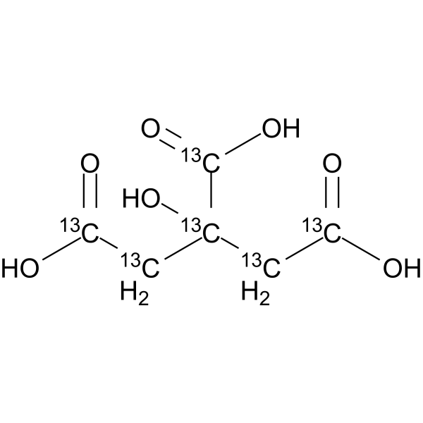
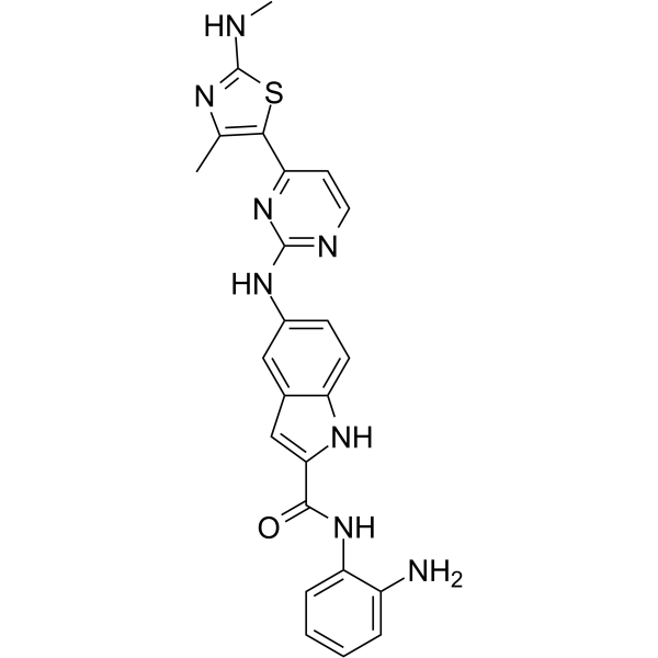
Topoisomerase II inhibitor 14 是一种强效的拓扑异构酶 II (topoisomerase II ) 抑制剂。Topoisomerase II inhibitor 14 可诱导细胞凋亡 (apoptosis),并将细胞周期阻滞于 S 期和 G2-M 期。Topoisomerase II inhibitor 14 具有抗氧化作用,可降低谷胱甘肽 (GSH)、丙二醛 (MDA) 和一氧化氮 (NO) 的水平。Topoisomerase II inhibitor 14 可用于颈部鳞状细胞癌 (HNSCC) 的研究。
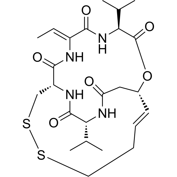
(S)-Seco-Duocarmycin SA 是 Seco-Duocarmycin SA (HY-129356) 的 S-对映体。Seco-Duocarmycin SA 是一种 DNA 烷化剂。Seco-Duocarmycin SA 是一种抗肿瘤抗生素 (IC50 = 10 pM)。Seco-Duocarmycin SA 可诱导凋亡细胞死亡 (apoptosis),且呈浓度依赖性增加。Seco-Duocarmycin SA 可导致细胞周期显著停滞于 S 期和 G2/M 期。Seco-Duocarmycin SA 可作为抗体-药物偶联物的 ADC 细胞毒素。
Seco-Duocarmycin SA 是一种 DNA 烷化剂 (DNA alkylator)。Seco-Duocarmycin SA 是一种抗肿瘤抗生素 (IC50 = 10 pM)。Seco-Duocarmycin SA 可诱导凋亡细胞死亡 (apoptosis),且呈浓度依赖性增加。Seco-Duocarmycin SA 可导致细胞周期显著停滞于 S 期和 G2/M 期。Seco-Duocarmycin SA 可作为抗体-药物偶联物的 ADC 细胞毒素。
CDK1; cyclin-dependent kinase 1; CDC2, cell division cycle 2, G1 to S and G2 to M; CDC28A; p34 protein kinase; cell cycle controller CDC2; cell division protein kinase 1; cell division control protein 2 homolog; cell division cycle 2, G1 to S and G2 to M; CDC2; P34CDC2; MGC111195; DKFZp686L20222
CDK1; cyclin-dependent kinase 1; CDC2, cell division cycle 2, G1 to S and G2 to M; CDC28A; p34 protein kinase; cell cycle controller CDC2; cell division protein kinase 1; cell division control protein 2 homolog; cell division cycle 2, G1 to S and G2 to M; CDC2; P34CDC2; MGC111195; DKFZp686L20222
CDK1; cyclin-dependent kinase 1; CDC2, cell division cycle 2, G1 to S and G2 to M; CDC28A; p34 protein kinase; cell cycle controller CDC2; cell division protein kinase 1; cell division control protein 2 homolog; cell division cycle 2, G1 to S and G2 to M; CDC2; P34CDC2; MGC111195; DKFZp686L20222
CDK1; cyclin-dependent kinase 1; CDC2, cell division cycle 2, G1 to S and G2 to M; CDC28A; p34 protein kinase; cell cycle controller CDC2; cell division protein kinase 1; cell division control protein 2 homolog; cell division cycle 2, G1 to S and G2 to M; CDC2; P34CDC2; MGC111195; DKFZp686L20222
Western blot analysis of extracts from THP-1(lane 2(20μg), Jurkat (lane 3(20μg) and NIH3T3(lane 4(20μg) using FOXO1A (HY-P80132) Rabbit mAb. Proteins were transferred
to a PVDF membrane and blocked with 5% non-fat milk in TBST for 2 hour at room temperature. The primary antibody (1/1000) and Loading control antibody (Beta Actin, HY-P80438, 1/10000) was
used in 5% non-fat milk in TBST at 4°C overnight. Goat Anti-Mouse/Rabbit IgG-HRP Secondary Antibody (1/10000) was used for 1 hour at room temperature.
Western blot analysis of extracts from THP-1(lane 2(20μg), Jurkat (lane 3(20μg) and NIH3T3(lane 4(20μg) using FOXO1A (HY-P80132) Rabbit mAb. Proteins were transferred
to a PVDF membrane and blocked with 5% non-fat milk in TBST for 2 hour at room temperature. The primary antibody (1/1000) and Loading control antibody (Beta Actin, HY-P80438, 1/10000) was
MedchemExpress Validation 03
Western blot analysis of extracts from THP-1(lane 2(20μg), Jurkat (lane 3(20μg) and NIH3T3(lane 4(20μg) using FOXO1A (HY-P80132) Rabbit mAb. Proteins were transferred
MedchemExpress Validation 04
Western blot analysis of extracts from THP-1(lane 2(20μg), Jurkat (lane 3(20μg) and NIH3T3(lane 4(20μg) using FOXO1A (HY-P80132) Rabbit mAb. Proteins were transferred
to a PVDF membrane and blocked with 5% non-fat milk in TBST for 2 hour at room temperature. The primary antibody (1/1000) and Loading control antibody (Beta Actin, HY-P80438, 1/10000) was
used in 5% non-fat milk in TBST at 4°C overnight. Goat Anti-Mouse/Rabbit IgG-HRP Secondary Antibody (1/10000) was used for 1 hour at room temperature.
MedchemExpress Validation
Western blot analysis of extracts from THP-1(lane 2(20μg), Jurkat (lane 3(20μg) and NIH3T3(lane 4(20μg) using FOXO1A (HY-P80132) Rabbit mAb. Proteins were transferred
to a PVDF membrane and blocked with 5% non-fat milk in TBST for 2 hour at room temperature. The primary antibody (1/1000) and Loading control antibody (Beta Actin, HY-P80438, 1/10000) was
used in 5% non-fat milk in TBST at 4°C overnight. Goat Anti-Mouse/Rabbit IgG-HRP Secondary Antibody (1/10000) was used for 1 hour at room temperature.
MedchemExpress Validation
Western blot analysis of extracts from THP-1(lane 2(20μg), Jurkat (lane 3(20μg) and NIH3T3(lane 4(20μg) using FOXO1A (HY-P80132) Rabbit mAb. Proteins were transferred
to a PVDF membrane and blocked with 5% non-fat milk in TBST for 2 hour at room temperature. The primary antibody (1/1000) and Loading control antibody (Beta Actin, HY-P80438, 1/10000) was
used in 5% non-fat milk in TBST at 4°C overnight. Goat Anti-Mouse/Rabbit IgG-HRP Secondary Antibody (1/10000) was used for 1 hour at room temperature.