Celliton Fast Blue Green B (Disperse Blue 7) 是一种用于纺织品的蓝绿色染料。Celliton Fast Blue Green B 的水提取物在实验动物中没有引起皮肤刺激和致敏的迹象。Celliton Fast Blue Green B 染色纺织品,对人体无刺激。
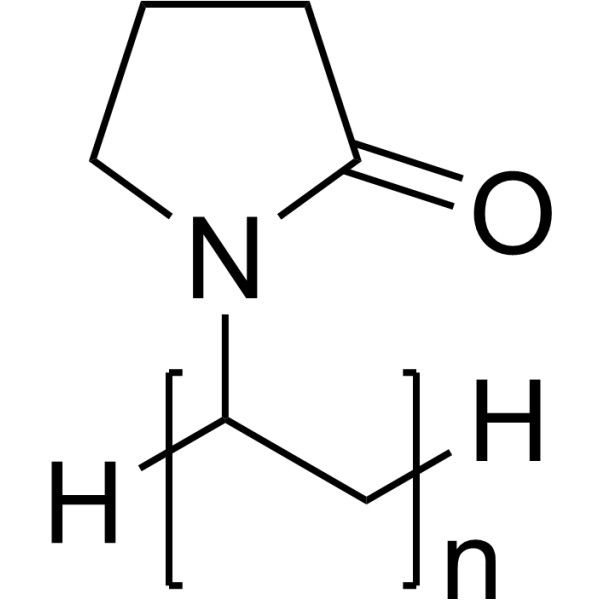
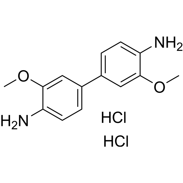
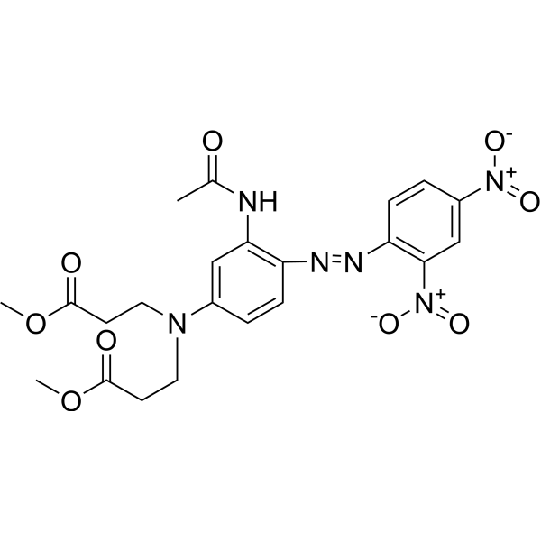
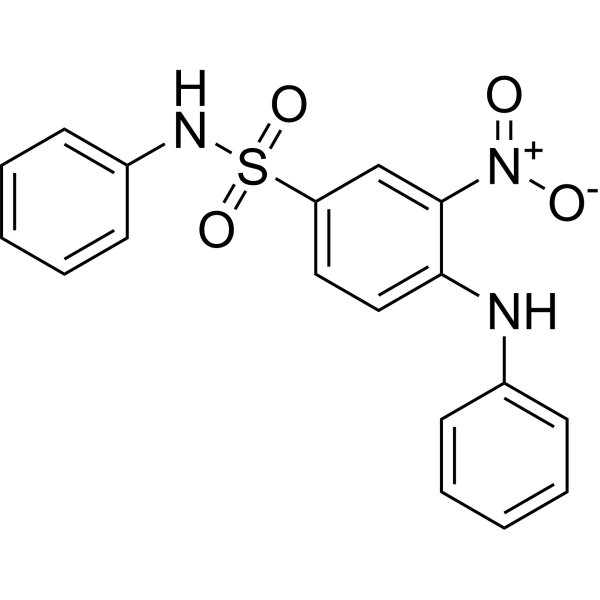
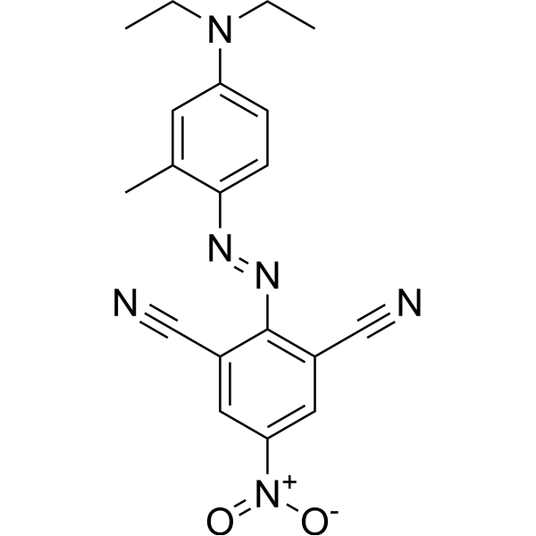
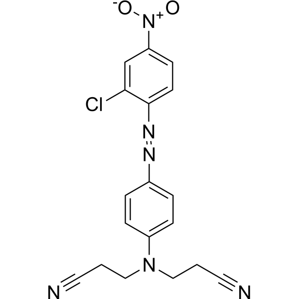
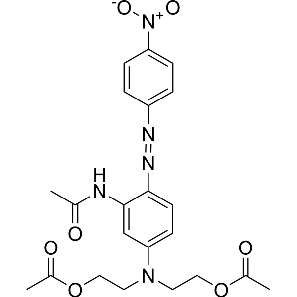
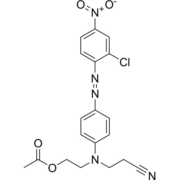
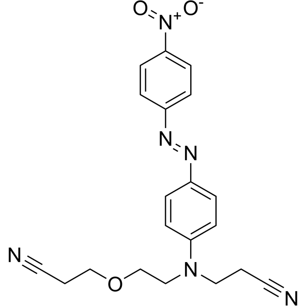
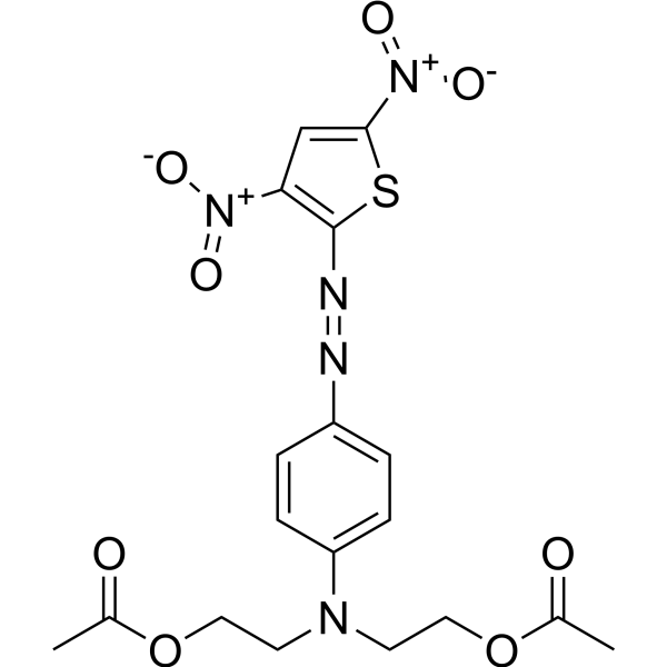
Disperse blue 291 是一种多功能染料。染料是生物实验中的重要工具,能帮助研究人员观察和分析细胞结构、追踪生物分子、评估细胞功能、区分细胞类型、检测生物分子、研究组织病理及监测微生物,应用涵盖从基础科学研究到临床诊断的广泛领域。染料在传统领域如纺织品染色,以及新兴领域如功能性纺织品处理、食品色素和染料敏化太阳能电池等,也有广泛应用。
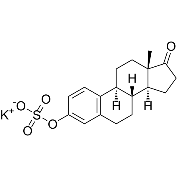
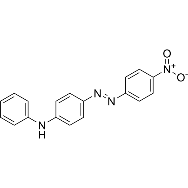
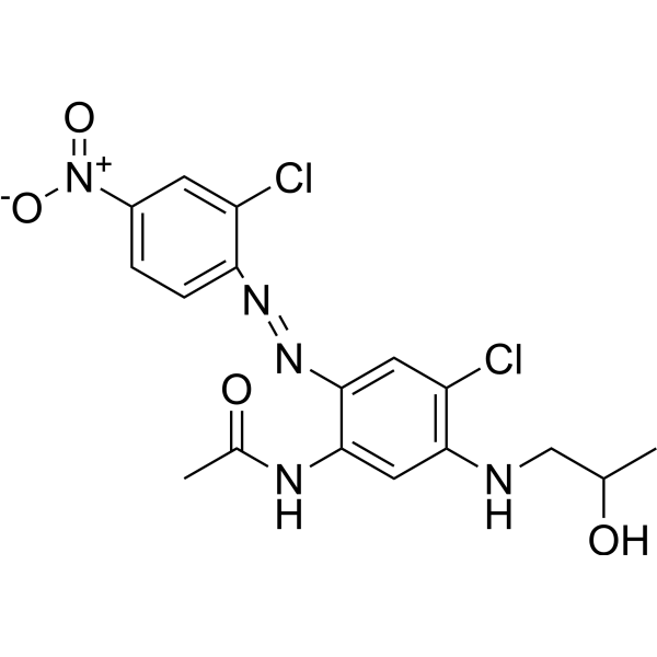
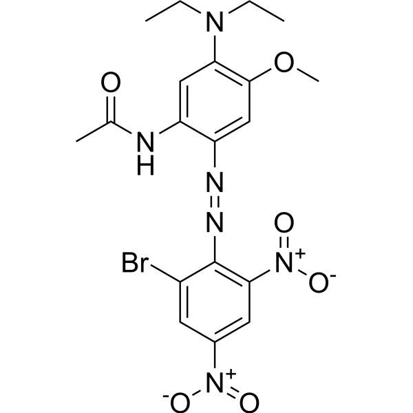
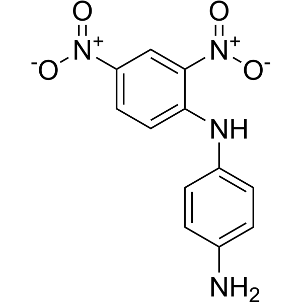
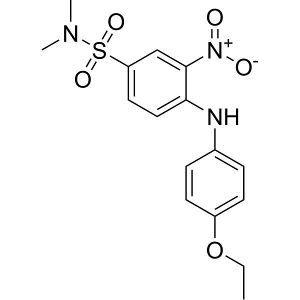
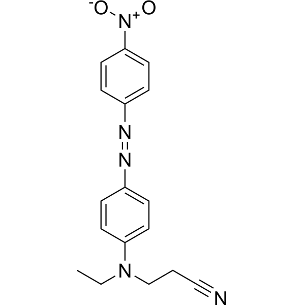
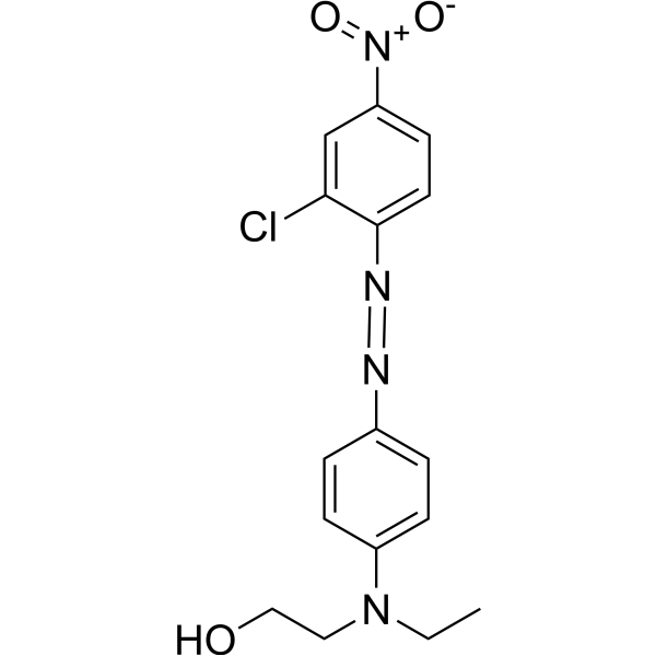
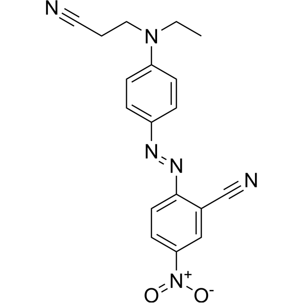
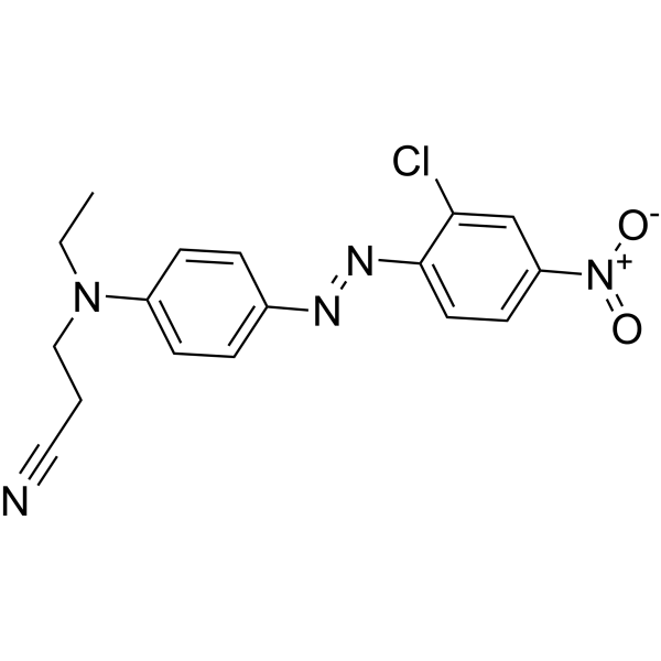
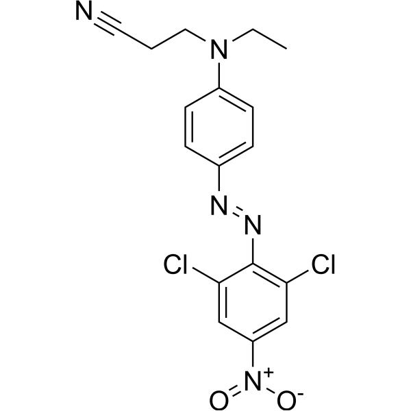
Disperse orange A 是一种多功能染料。染料是生物实验中的重要工具,能帮助研究人员观察和分析细胞结构、追踪生物分子、评估细胞功能、区分细胞类型、检测生物分子、研究组织病理及监测微生物,应用涵盖从基础科学研究到临床诊断的广泛领域。染料在传统领域如纺织品染色,以及新兴领域如功能性纺织品处理、食品色素和染料敏化太阳能电池等,也有广泛应用。
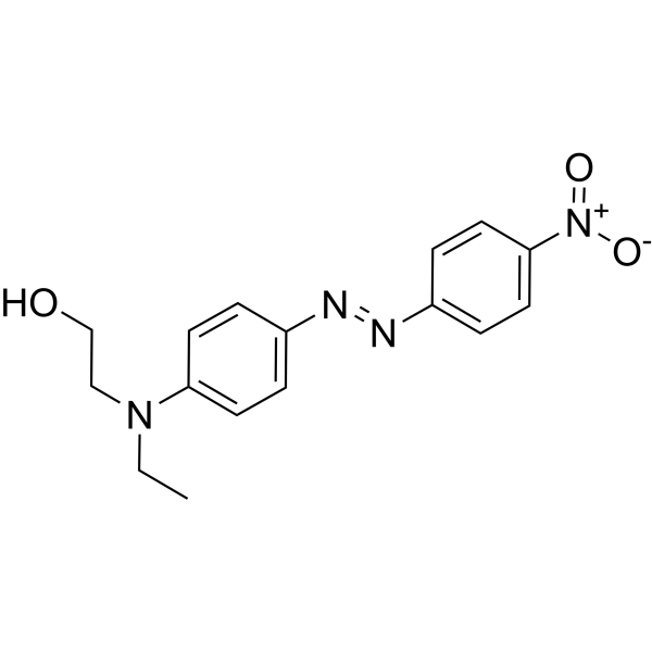
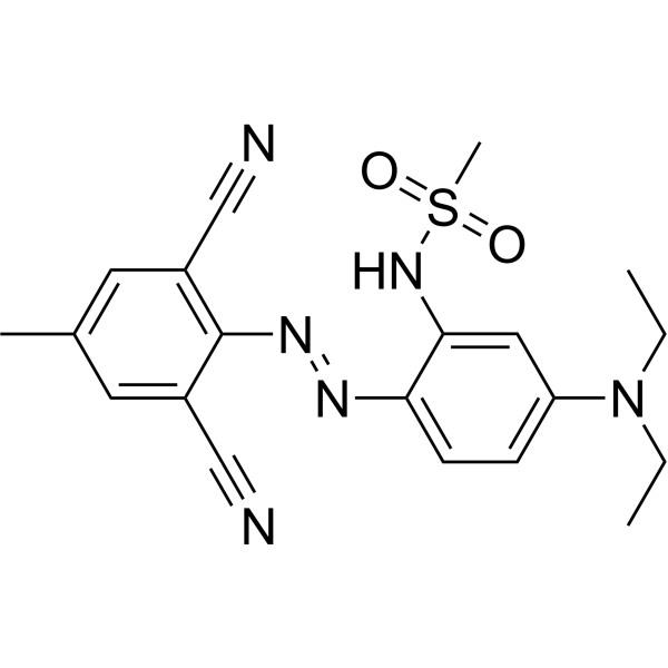
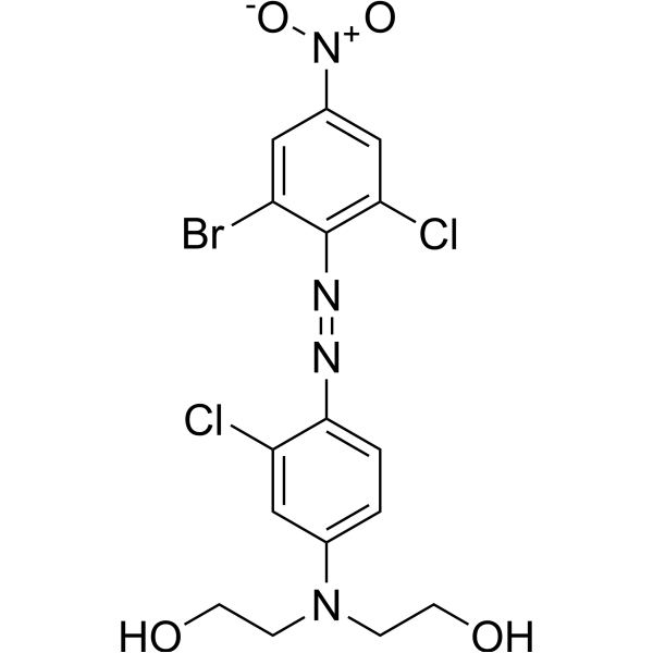
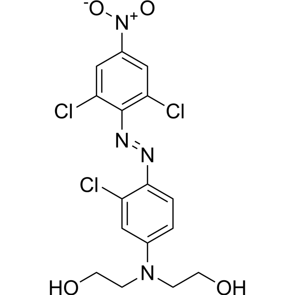
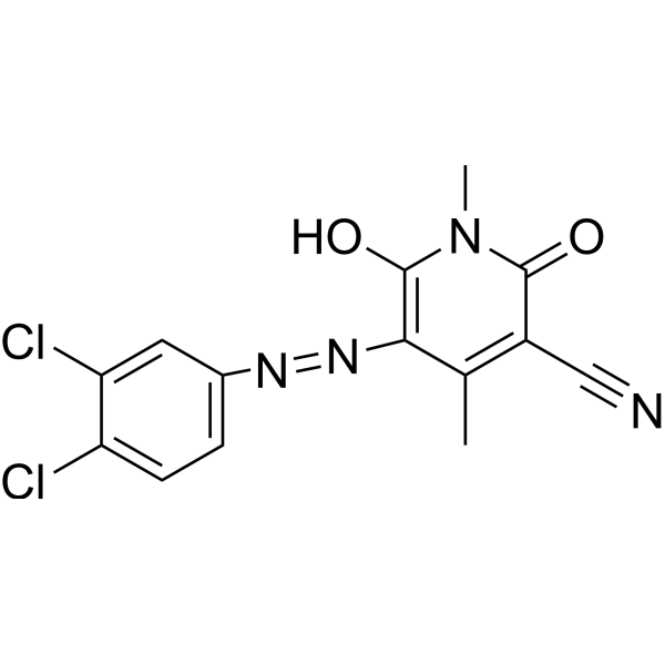
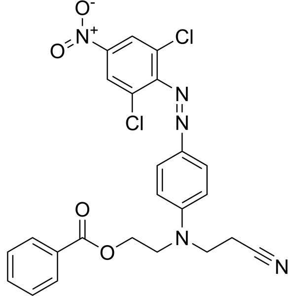
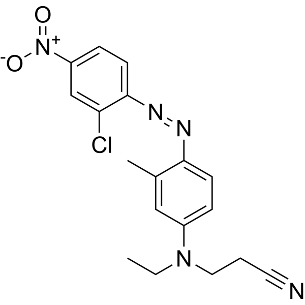
Disperse blue 165 是一种多功能染料。染料是生物实验中的重要工具,能帮助研究人员观察和分析细胞结构、追踪生物分子、评估细胞功能、区分细胞类型、检测生物分子、研究组织病理及监测微生物,应用涵盖从基础科学研究到临床诊断的广泛领域。染料在传统领域如纺织品染色,以及新兴领域如功能性纺织品处理、食品色素和染料敏化太阳能电池等,也有广泛应用。
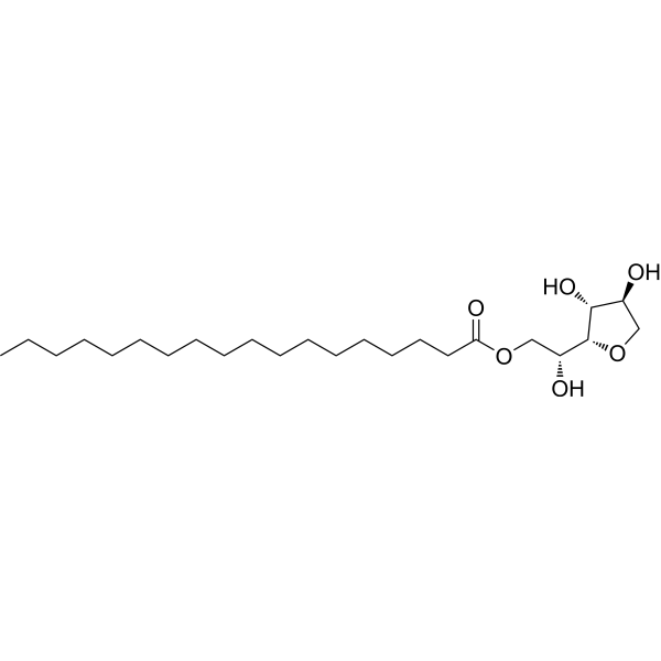
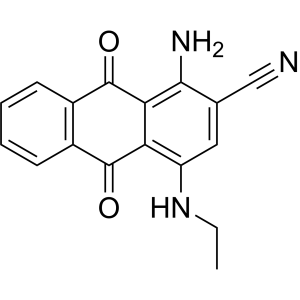
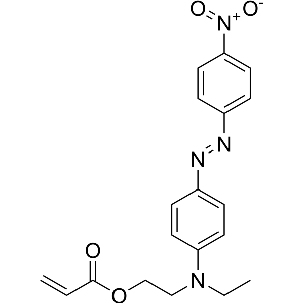
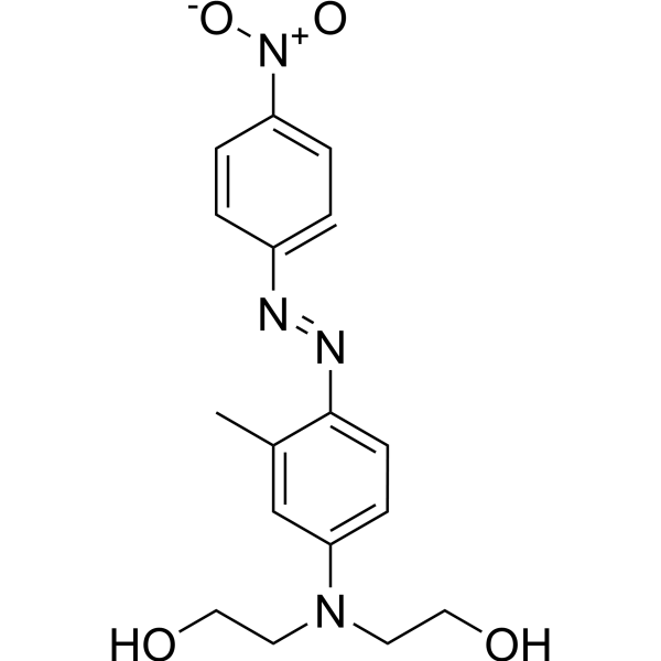
Disperse green 9 是一种多功能染料。染料是生物实验中的重要工具,能帮助研究人员观察和分析细胞结构、追踪生物分子、评估细胞功能、区分细胞类型、检测生物分子、研究组织病理及监测微生物,应用涵盖从基础科学研究到临床诊断的广泛领域。染料在传统领域如纺织品染色,以及新兴领域如功能性纺织品处理、食品色素和染料敏化太阳能电池等,也有广泛应用。
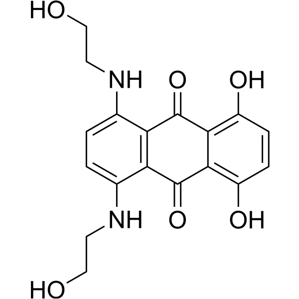
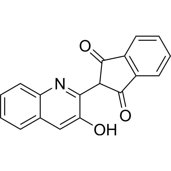
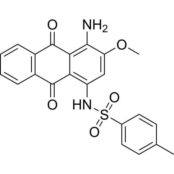
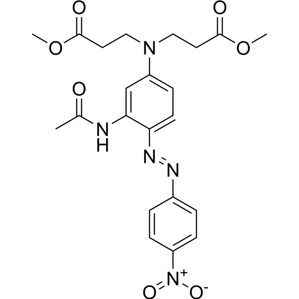
C.I. Disperse blue 284 是一种多功能染料。染料是生物实验中的重要工具,能帮助研究人员观察和分析细胞结构、追踪生物分子、评估细胞功能、区分细胞类型、检测生物分子、研究组织病理及监测微生物,应用涵盖从基础科学研究到临床诊断的广泛领域。染料在传统领域如纺织品染色,以及新兴领域如功能性纺织品处理、食品色素和染料敏化太阳能电池等,也有广泛应用。
Disperse blue 291 是一种多功能染料。染料是生物实验中的重要工具,能帮助研究人员观察和分析细胞结构、追踪生物分子、评估细胞功能、区分细胞类型、检测生物分子、研究组织病理及监测微生物,应用涵盖从基础科学研究到临床诊断的广泛领域。染料在传统领域如纺织品染色,以及新兴领域如功能性纺织品处理、食品色素和染料敏化太阳能电池等,也有广泛应用。
Disperse orange A 是一种多功能染料。染料是生物实验中的重要工具,能帮助研究人员观察和分析细胞结构、追踪生物分子、评估细胞功能、区分细胞类型、检测生物分子、研究组织病理及监测微生物,应用涵盖从基础科学研究到临床诊断的广泛领域。染料在传统领域如纺织品染色,以及新兴领域如功能性纺织品处理、食品色素和染料敏化太阳能电池等,也有广泛应用。
Disperse blue 165 是一种多功能染料。染料是生物实验中的重要工具,能帮助研究人员观察和分析细胞结构、追踪生物分子、评估细胞功能、区分细胞类型、检测生物分子、研究组织病理及监测微生物,应用涵盖从基础科学研究到临床诊断的广泛领域。染料在传统领域如纺织品染色,以及新兴领域如功能性纺织品处理、食品色素和染料敏化太阳能电池等,也有广泛应用。
Disperse green 9 是一种多功能染料。染料是生物实验中的重要工具,能帮助研究人员观察和分析细胞结构、追踪生物分子、评估细胞功能、区分细胞类型、检测生物分子、研究组织病理及监测微生物,应用涵盖从基础科学研究到临床诊断的广泛领域。染料在传统领域如纺织品染色,以及新兴领域如功能性纺织品处理、食品色素和染料敏化太阳能电池等,也有广泛应用。
C.I. Disperse blue 284 是一种多功能染料。染料是生物实验中的重要工具,能帮助研究人员观察和分析细胞结构、追踪生物分子、评估细胞功能、区分细胞类型、检测生物分子、研究组织病理及监测微生物,应用涵盖从基础科学研究到临床诊断的广泛领域。染料在传统领域如纺织品染色,以及新兴领域如功能性纺织品处理、食品色素和染料敏化太阳能电池等,也有广泛应用。
Western blot analysis of extracts from THP-1(lane 2(20μg), Jurkat (lane 3(20μg) and NIH3T3(lane 4(20μg) using FOXO1A (HY-P80132) Rabbit mAb. Proteins were transferred
to a PVDF membrane and blocked with 5% non-fat milk in TBST for 2 hour at room temperature. The primary antibody (1/1000) and Loading control antibody (Beta Actin, HY-P80438, 1/10000) was
used in 5% non-fat milk in TBST at 4°C overnight. Goat Anti-Mouse/Rabbit IgG-HRP Secondary Antibody (1/10000) was used for 1 hour at room temperature.
Western blot analysis of extracts from THP-1(lane 2(20μg), Jurkat (lane 3(20μg) and NIH3T3(lane 4(20μg) using FOXO1A (HY-P80132) Rabbit mAb. Proteins were transferred
to a PVDF membrane and blocked with 5% non-fat milk in TBST for 2 hour at room temperature. The primary antibody (1/1000) and Loading control antibody (Beta Actin, HY-P80438, 1/10000) was
MedchemExpress Validation 03
Western blot analysis of extracts from THP-1(lane 2(20μg), Jurkat (lane 3(20μg) and NIH3T3(lane 4(20μg) using FOXO1A (HY-P80132) Rabbit mAb. Proteins were transferred
MedchemExpress Validation 04
Western blot analysis of extracts from THP-1(lane 2(20μg), Jurkat (lane 3(20μg) and NIH3T3(lane 4(20μg) using FOXO1A (HY-P80132) Rabbit mAb. Proteins were transferred
to a PVDF membrane and blocked with 5% non-fat milk in TBST for 2 hour at room temperature. The primary antibody (1/1000) and Loading control antibody (Beta Actin, HY-P80438, 1/10000) was
used in 5% non-fat milk in TBST at 4°C overnight. Goat Anti-Mouse/Rabbit IgG-HRP Secondary Antibody (1/10000) was used for 1 hour at room temperature.
MedchemExpress Validation
Western blot analysis of extracts from THP-1(lane 2(20μg), Jurkat (lane 3(20μg) and NIH3T3(lane 4(20μg) using FOXO1A (HY-P80132) Rabbit mAb. Proteins were transferred
to a PVDF membrane and blocked with 5% non-fat milk in TBST for 2 hour at room temperature. The primary antibody (1/1000) and Loading control antibody (Beta Actin, HY-P80438, 1/10000) was
used in 5% non-fat milk in TBST at 4°C overnight. Goat Anti-Mouse/Rabbit IgG-HRP Secondary Antibody (1/10000) was used for 1 hour at room temperature.
MedchemExpress Validation
Western blot analysis of extracts from THP-1(lane 2(20μg), Jurkat (lane 3(20μg) and NIH3T3(lane 4(20μg) using FOXO1A (HY-P80132) Rabbit mAb. Proteins were transferred
to a PVDF membrane and blocked with 5% non-fat milk in TBST for 2 hour at room temperature. The primary antibody (1/1000) and Loading control antibody (Beta Actin, HY-P80438, 1/10000) was
used in 5% non-fat milk in TBST at 4°C overnight. Goat Anti-Mouse/Rabbit IgG-HRP Secondary Antibody (1/10000) was used for 1 hour at room temperature.