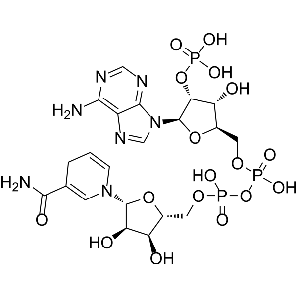
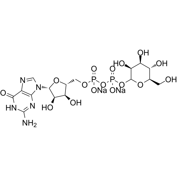
糖苷内切酶 D
Endo-β-N-acetylglucosaminidase D (Endo D) 能够从肺炎链球菌 (Streptococcus pneumoniae) 中分离得到。在连续去除 N-聚糖中的唾液酸、半乳糖和内部 GlcNAc 残基后,Endo-β-N-acetylglucosaminidase D 可水解完整 IgG 抗体的 Fc N-聚糖。Endo-β-N-acetylglucosaminidase D 具有以糖恶唑啉为供体底物的转糖基活性。
Cathepsin E substrate e 是组织蛋白酶 E 的底物。Cathepsin E substrate e 的设计方式是,由于 Mca 供体和 Dnp 受体非常接近,因此在完整状态下可实现近乎完全的分子内猝灭效应。肽底物的疏水基序被蛋白酶切割后,Mca 和 Dnp 会进一步分离,从而产生明亮的荧光。
Oxalate oxidase, B. subtilis (EC 1.2.3.4) 是一种作用于供体醛基或羰基,以氧为受体的氧化还原酶。Oxalate oxidase, B 参与乙醛酸和二羧酸的代谢。Oxalate oxidase, B 有两种辅因子:FAD 和锰。Oxalate oxidase, B 的三种底物是草酸、O2 和 H+,而其两种产物是 CO2 和 H2O2。
Cathepsin E substrate e 是组织蛋白酶 E 的底物。Cathepsin E substrate e 的设计方式是,由于 Mca 供体和 Dnp 受体非常接近,因此在完整状态下可实现近乎完全的分子内猝灭效应。肽底物的疏水基序被蛋白酶切割后,Mca 和 Dnp 会进一步分离,从而产生明亮的荧光。
Western blot analysis of extracts from THP-1(lane 2(20μg), Jurkat (lane 3(20μg) and NIH3T3(lane 4(20μg) using FOXO1A (HY-P80132) Rabbit mAb. Proteins were transferred
to a PVDF membrane and blocked with 5% non-fat milk in TBST for 2 hour at room temperature. The primary antibody (1/1000) and Loading control antibody (Beta Actin, HY-P80438, 1/10000) was
used in 5% non-fat milk in TBST at 4°C overnight. Goat Anti-Mouse/Rabbit IgG-HRP Secondary Antibody (1/10000) was used for 1 hour at room temperature.
Western blot analysis of extracts from THP-1(lane 2(20μg), Jurkat (lane 3(20μg) and NIH3T3(lane 4(20μg) using FOXO1A (HY-P80132) Rabbit mAb. Proteins were transferred
to a PVDF membrane and blocked with 5% non-fat milk in TBST for 2 hour at room temperature. The primary antibody (1/1000) and Loading control antibody (Beta Actin, HY-P80438, 1/10000) was
MedchemExpress Validation 03
Western blot analysis of extracts from THP-1(lane 2(20μg), Jurkat (lane 3(20μg) and NIH3T3(lane 4(20μg) using FOXO1A (HY-P80132) Rabbit mAb. Proteins were transferred
MedchemExpress Validation 04
Western blot analysis of extracts from THP-1(lane 2(20μg), Jurkat (lane 3(20μg) and NIH3T3(lane 4(20μg) using FOXO1A (HY-P80132) Rabbit mAb. Proteins were transferred
to a PVDF membrane and blocked with 5% non-fat milk in TBST for 2 hour at room temperature. The primary antibody (1/1000) and Loading control antibody (Beta Actin, HY-P80438, 1/10000) was
used in 5% non-fat milk in TBST at 4°C overnight. Goat Anti-Mouse/Rabbit IgG-HRP Secondary Antibody (1/10000) was used for 1 hour at room temperature.
MedchemExpress Validation
Western blot analysis of extracts from THP-1(lane 2(20μg), Jurkat (lane 3(20μg) and NIH3T3(lane 4(20μg) using FOXO1A (HY-P80132) Rabbit mAb. Proteins were transferred
to a PVDF membrane and blocked with 5% non-fat milk in TBST for 2 hour at room temperature. The primary antibody (1/1000) and Loading control antibody (Beta Actin, HY-P80438, 1/10000) was
used in 5% non-fat milk in TBST at 4°C overnight. Goat Anti-Mouse/Rabbit IgG-HRP Secondary Antibody (1/10000) was used for 1 hour at room temperature.
MedchemExpress Validation
Western blot analysis of extracts from THP-1(lane 2(20μg), Jurkat (lane 3(20μg) and NIH3T3(lane 4(20μg) using FOXO1A (HY-P80132) Rabbit mAb. Proteins were transferred
to a PVDF membrane and blocked with 5% non-fat milk in TBST for 2 hour at room temperature. The primary antibody (1/1000) and Loading control antibody (Beta Actin, HY-P80438, 1/10000) was
used in 5% non-fat milk in TBST at 4°C overnight. Goat Anti-Mouse/Rabbit IgG-HRP Secondary Antibody (1/10000) was used for 1 hour at room temperature.