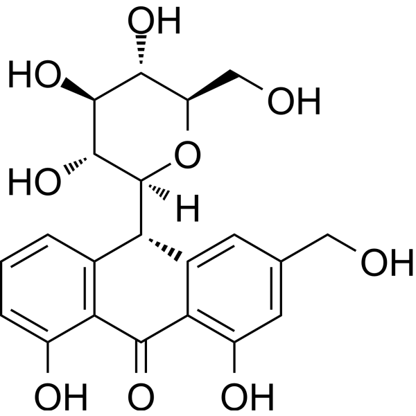
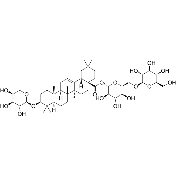
川续断皂苷 VI
Asperosaponin VI 是来自川续断的一种皂苷组分。Asperosaponin VI 通过 BMP-2/p38 和 ERK1/2 信号途径诱导成骨细胞分化。Asperosaponin VI 通过激活 PI3K/Akt 和 CREB 通路保护缺氧诱导的心肌细胞凋亡 (apoptosis)。此外,Asperosaponin VI 还具有抗抑郁和促伤口愈合的活性。
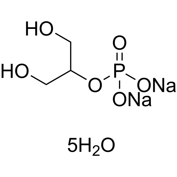
2,3-Diphospho-D-glyceric acid (2,3-DPG) pentasodium salt 是一种血红蛋白结合剂和血管钙化抑制剂,可降低血红蛋白的氧亲和力。2,3-Diphospho-D-glyceric acid pentasodium salt 还能特异性地延缓胶体钙蛋白颗粒向结晶型转化,从而有效抑制血管平滑肌细胞钙化,且不影响成骨样细胞中骨样结节的正常形成。2,3-Diphospho-D-glyceric acid pentasodium salt 对受试细胞系无细胞毒性,且对单棕榈酸甘油介导的 β-血红素形成仅具微弱干扰。2,3-Diphospho-D-glyceric acid pentasodium salt 可用于研究血管钙化病理机制和疟疾相关。
川续断皂苷 VI
Asperosaponin VI 是来自川续断的一种皂苷组分。Asperosaponin VI 通过 BMP-2/p38 和 ERK1/2 信号途径诱导成骨细胞分化。Asperosaponin VI 通过激活 PI3K/Akt 和 CREB 通路保护缺氧诱导的心肌细胞凋亡 (apoptosis)。此外,Asperosaponin VI 还具有抗抑郁和促伤口愈合的活性。
Western blot analysis of extracts from THP-1(lane 2(20μg), Jurkat (lane 3(20μg) and NIH3T3(lane 4(20μg) using FOXO1A (HY-P80132) Rabbit mAb. Proteins were transferred
to a PVDF membrane and blocked with 5% non-fat milk in TBST for 2 hour at room temperature. The primary antibody (1/1000) and Loading control antibody (Beta Actin, HY-P80438, 1/10000) was
used in 5% non-fat milk in TBST at 4°C overnight. Goat Anti-Mouse/Rabbit IgG-HRP Secondary Antibody (1/10000) was used for 1 hour at room temperature.
Western blot analysis of extracts from THP-1(lane 2(20μg), Jurkat (lane 3(20μg) and NIH3T3(lane 4(20μg) using FOXO1A (HY-P80132) Rabbit mAb. Proteins were transferred
to a PVDF membrane and blocked with 5% non-fat milk in TBST for 2 hour at room temperature. The primary antibody (1/1000) and Loading control antibody (Beta Actin, HY-P80438, 1/10000) was
MedchemExpress Validation 03
Western blot analysis of extracts from THP-1(lane 2(20μg), Jurkat (lane 3(20μg) and NIH3T3(lane 4(20μg) using FOXO1A (HY-P80132) Rabbit mAb. Proteins were transferred
MedchemExpress Validation 04
Western blot analysis of extracts from THP-1(lane 2(20μg), Jurkat (lane 3(20μg) and NIH3T3(lane 4(20μg) using FOXO1A (HY-P80132) Rabbit mAb. Proteins were transferred
to a PVDF membrane and blocked with 5% non-fat milk in TBST for 2 hour at room temperature. The primary antibody (1/1000) and Loading control antibody (Beta Actin, HY-P80438, 1/10000) was
used in 5% non-fat milk in TBST at 4°C overnight. Goat Anti-Mouse/Rabbit IgG-HRP Secondary Antibody (1/10000) was used for 1 hour at room temperature.
MedchemExpress Validation
Western blot analysis of extracts from THP-1(lane 2(20μg), Jurkat (lane 3(20μg) and NIH3T3(lane 4(20μg) using FOXO1A (HY-P80132) Rabbit mAb. Proteins were transferred
to a PVDF membrane and blocked with 5% non-fat milk in TBST for 2 hour at room temperature. The primary antibody (1/1000) and Loading control antibody (Beta Actin, HY-P80438, 1/10000) was
used in 5% non-fat milk in TBST at 4°C overnight. Goat Anti-Mouse/Rabbit IgG-HRP Secondary Antibody (1/10000) was used for 1 hour at room temperature.
MedchemExpress Validation
Western blot analysis of extracts from THP-1(lane 2(20μg), Jurkat (lane 3(20μg) and NIH3T3(lane 4(20μg) using FOXO1A (HY-P80132) Rabbit mAb. Proteins were transferred
to a PVDF membrane and blocked with 5% non-fat milk in TBST for 2 hour at room temperature. The primary antibody (1/1000) and Loading control antibody (Beta Actin, HY-P80438, 1/10000) was
used in 5% non-fat milk in TBST at 4°C overnight. Goat Anti-Mouse/Rabbit IgG-HRP Secondary Antibody (1/10000) was used for 1 hour at room temperature.