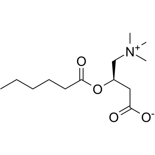
Avenanthramide A 是一种植物防御素,存在于燕麦 (Avena sativa L.) 中。Avenanthramide A 靶向 RNA 解旋酶 DDX3,导致线粒体肿胀和 ROS 生成增加,并诱导 CRC 细胞凋亡 (apoptosis)。Avenanthramide A 在小鼠模型中表现出抗肿瘤活性。Avenanthramide A 具有口服活性。
甘草异黄酮 A
Licoisoflavone A 是一种具有口服活性的异黄酮。Licoisoflavone A 抑制结直肠癌 (CRC) 细胞的增殖,诱导凋亡 (apoptosis),并引起 G1/S 期阻滞。Licoisoflavone A 抑制 CDK2-CyclinE1 轴。Licoisoflavone A 抑制脂质过氧化,IC50 为 7.2 μM。Licoisoflavone A 对 SARS-CoV-2 感染表现出剂量依赖的抑制效果。Licoisoflavone A 在携带 CT26 细胞皮下异种移植的小鼠中显示出显著的抗肿瘤效能。Licoisoflavone A 可用于结直肠癌和 SARS-CoV-2 感染研究。
Avenanthramide A (Standard)是 Avenanthramide A 的分析标准品。本产品用于研究及分析应用。Avenanthramide A 是一种植物防御素,存在于燕麦 (Avena sativa L.) 中。Avenanthramide A 靶向 RNA 解旋酶 DDX3,导致线粒体肿胀和 ROS 生成增加,并诱导 CRC 细胞凋亡 (apoptosis)。Avenanthramide A 在小鼠模型中表现出抗肿瘤活性。Avenanthramide A 具有口服活性。
Avenanthramide A-d4 是氘标记的 Avenanthramide A (HY-114977)。Avenanthramide A 是一种植物防御素,可在燕麦 (Avena sativa L.) 中发现。Avenanthramide A 靶向 RNA 解旋酶 DDX3,导致线粒体肿胀和 ROS 生成增加,并诱导 CRC 细胞凋亡 (apoptosis)。Avenanthramide A 在小鼠模型中表现出抗肿瘤活性。Avenanthramide A 具有口服活性。
Western blot analysis of extracts from THP-1(lane 2(20μg), Jurkat (lane 3(20μg) and NIH3T3(lane 4(20μg) using FOXO1A (HY-P80132) Rabbit mAb. Proteins were transferred
to a PVDF membrane and blocked with 5% non-fat milk in TBST for 2 hour at room temperature. The primary antibody (1/1000) and Loading control antibody (Beta Actin, HY-P80438, 1/10000) was
used in 5% non-fat milk in TBST at 4°C overnight. Goat Anti-Mouse/Rabbit IgG-HRP Secondary Antibody (1/10000) was used for 1 hour at room temperature.
Western blot analysis of extracts from THP-1(lane 2(20μg), Jurkat (lane 3(20μg) and NIH3T3(lane 4(20μg) using FOXO1A (HY-P80132) Rabbit mAb. Proteins were transferred
to a PVDF membrane and blocked with 5% non-fat milk in TBST for 2 hour at room temperature. The primary antibody (1/1000) and Loading control antibody (Beta Actin, HY-P80438, 1/10000) was
MedchemExpress Validation 03
Western blot analysis of extracts from THP-1(lane 2(20μg), Jurkat (lane 3(20μg) and NIH3T3(lane 4(20μg) using FOXO1A (HY-P80132) Rabbit mAb. Proteins were transferred
MedchemExpress Validation 04
Western blot analysis of extracts from THP-1(lane 2(20μg), Jurkat (lane 3(20μg) and NIH3T3(lane 4(20μg) using FOXO1A (HY-P80132) Rabbit mAb. Proteins were transferred
to a PVDF membrane and blocked with 5% non-fat milk in TBST for 2 hour at room temperature. The primary antibody (1/1000) and Loading control antibody (Beta Actin, HY-P80438, 1/10000) was
used in 5% non-fat milk in TBST at 4°C overnight. Goat Anti-Mouse/Rabbit IgG-HRP Secondary Antibody (1/10000) was used for 1 hour at room temperature.
MedchemExpress Validation
Western blot analysis of extracts from THP-1(lane 2(20μg), Jurkat (lane 3(20μg) and NIH3T3(lane 4(20μg) using FOXO1A (HY-P80132) Rabbit mAb. Proteins were transferred
to a PVDF membrane and blocked with 5% non-fat milk in TBST for 2 hour at room temperature. The primary antibody (1/1000) and Loading control antibody (Beta Actin, HY-P80438, 1/10000) was
used in 5% non-fat milk in TBST at 4°C overnight. Goat Anti-Mouse/Rabbit IgG-HRP Secondary Antibody (1/10000) was used for 1 hour at room temperature.
MedchemExpress Validation
Western blot analysis of extracts from THP-1(lane 2(20μg), Jurkat (lane 3(20μg) and NIH3T3(lane 4(20μg) using FOXO1A (HY-P80132) Rabbit mAb. Proteins were transferred
to a PVDF membrane and blocked with 5% non-fat milk in TBST for 2 hour at room temperature. The primary antibody (1/1000) and Loading control antibody (Beta Actin, HY-P80438, 1/10000) was
used in 5% non-fat milk in TBST at 4°C overnight. Goat Anti-Mouse/Rabbit IgG-HRP Secondary Antibody (1/10000) was used for 1 hour at room temperature.


![[Ru(phen)2(xant)] hexafluorophosphate](http://hg.y866.cn/biomolecule/lib/file/product_pic/hy-161102.gif)