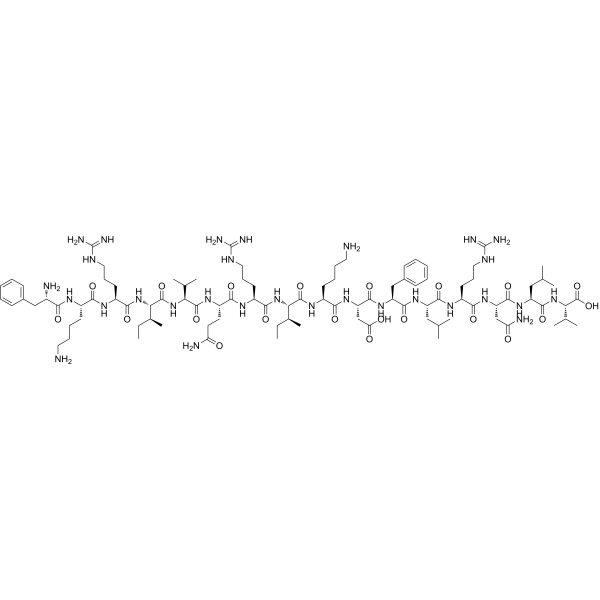
Plantaricin A 是一种抗菌肽,可从植物乳杆菌 (Lactobacillus plantarum) 中提取。Plantaricin A 与环丙沙星具有协同作用。Plantaricin A 表现出抗菌活性。Plantaricin A 可以提高金黄色葡萄球菌的膜电位和细胞内活性氧 (ROS) 水平。Plantaricin A 通过与外排泵结合并改变 MepA、NorA 和 LmrS 的结构,从而抑制外排泵的功能。Plantaricin A 能显著缓解炎症,促进伤口愈合。Plantaricin A 对癌变大鼠垂体细胞具有通透化作用[1][2]。
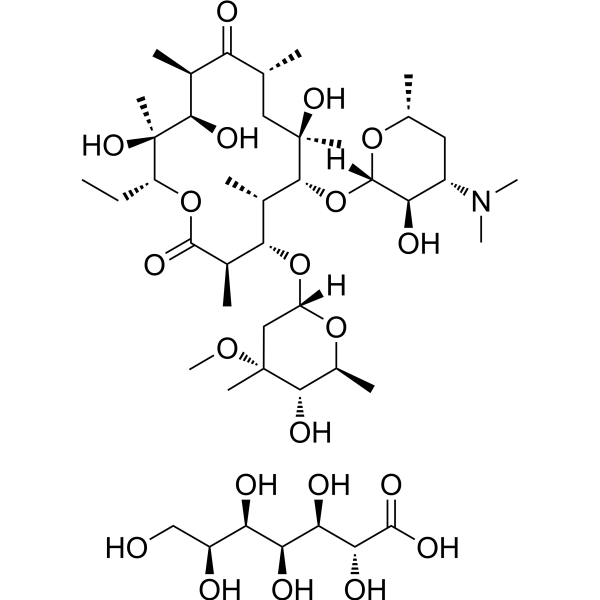
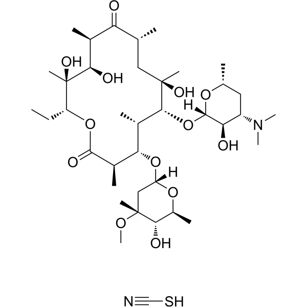
Erythromycin A dihydrate 是由放线菌产生的大环内酯类抗生素 (antibiotic),具有广泛的抗菌活性。Erythromycin A dihydrate 通过结合细菌的 50S 核糖体亚基而起作用,并通过阻断转肽和/或易位反应来抑制 RNA 依赖性蛋白 (RNA-dependent protein synthesis) 的合成,而不会影响核酸的合成。Erythromycin A dihydrate 在不同领域的研究中显示出抗肿瘤特性和保护神经的作用。
Plantaricin A 是一种抗菌肽,可从植物乳杆菌 (Lactobacillus plantarum) 中提取。Plantaricin A 与环丙沙星具有协同作用。Plantaricin A 表现出抗菌活性。Plantaricin A 可以提高金黄色葡萄球菌的膜电位和细胞内活性氧 (ROS) 水平。Plantaricin A 通过与外排泵结合并改变 MepA、NorA 和 LmrS 的结构,从而抑制外排泵的功能。Plantaricin A 能显著缓解炎症,促进伤口愈合。Plantaricin A 对癌变大鼠垂体细胞具有通透化作用[1][2]。
Western blot analysis of extracts from THP-1(lane 2(20μg), Jurkat (lane 3(20μg) and NIH3T3(lane 4(20μg) using FOXO1A (HY-P80132) Rabbit mAb. Proteins were transferred
to a PVDF membrane and blocked with 5% non-fat milk in TBST for 2 hour at room temperature. The primary antibody (1/1000) and Loading control antibody (Beta Actin, HY-P80438, 1/10000) was
used in 5% non-fat milk in TBST at 4°C overnight. Goat Anti-Mouse/Rabbit IgG-HRP Secondary Antibody (1/10000) was used for 1 hour at room temperature.
Western blot analysis of extracts from THP-1(lane 2(20μg), Jurkat (lane 3(20μg) and NIH3T3(lane 4(20μg) using FOXO1A (HY-P80132) Rabbit mAb. Proteins were transferred
to a PVDF membrane and blocked with 5% non-fat milk in TBST for 2 hour at room temperature. The primary antibody (1/1000) and Loading control antibody (Beta Actin, HY-P80438, 1/10000) was
MedchemExpress Validation 03
Western blot analysis of extracts from THP-1(lane 2(20μg), Jurkat (lane 3(20μg) and NIH3T3(lane 4(20μg) using FOXO1A (HY-P80132) Rabbit mAb. Proteins were transferred
MedchemExpress Validation 04
Western blot analysis of extracts from THP-1(lane 2(20μg), Jurkat (lane 3(20μg) and NIH3T3(lane 4(20μg) using FOXO1A (HY-P80132) Rabbit mAb. Proteins were transferred
to a PVDF membrane and blocked with 5% non-fat milk in TBST for 2 hour at room temperature. The primary antibody (1/1000) and Loading control antibody (Beta Actin, HY-P80438, 1/10000) was
used in 5% non-fat milk in TBST at 4°C overnight. Goat Anti-Mouse/Rabbit IgG-HRP Secondary Antibody (1/10000) was used for 1 hour at room temperature.
MedchemExpress Validation
Western blot analysis of extracts from THP-1(lane 2(20μg), Jurkat (lane 3(20μg) and NIH3T3(lane 4(20μg) using FOXO1A (HY-P80132) Rabbit mAb. Proteins were transferred
to a PVDF membrane and blocked with 5% non-fat milk in TBST for 2 hour at room temperature. The primary antibody (1/1000) and Loading control antibody (Beta Actin, HY-P80438, 1/10000) was
used in 5% non-fat milk in TBST at 4°C overnight. Goat Anti-Mouse/Rabbit IgG-HRP Secondary Antibody (1/10000) was used for 1 hour at room temperature.
MedchemExpress Validation
Western blot analysis of extracts from THP-1(lane 2(20μg), Jurkat (lane 3(20μg) and NIH3T3(lane 4(20μg) using FOXO1A (HY-P80132) Rabbit mAb. Proteins were transferred
to a PVDF membrane and blocked with 5% non-fat milk in TBST for 2 hour at room temperature. The primary antibody (1/1000) and Loading control antibody (Beta Actin, HY-P80438, 1/10000) was
used in 5% non-fat milk in TBST at 4°C overnight. Goat Anti-Mouse/Rabbit IgG-HRP Secondary Antibody (1/10000) was used for 1 hour at room temperature.