Cardiotoxin Analog (CTX) IV (6-12) 是一种特异性靶向带负电荷磷脂膜 (如磷脂酰丝氨酸、磷脂酰肌醇) 的膜活性肽段,能够从台湾眼镜蛇的毒液中分离得到。Cardiotoxin Analog (CTX) IV (6-12) 是一种蛇毒心脏毒素,通过疏水相互作用和静电吸引结合细胞膜、嵌入脂质双层,从而破坏膜结构稳定性。Cardiotoxin Analog (CTX) IV (6-12) 可诱导膜脂紊乱和细胞裂解,展现出溶血和细胞毒性。
Cardiotoxin Analog (CTX) IV (6-12) TFA 是一种特异性靶向带负电荷磷脂膜 (如磷脂酰丝氨酸、磷脂酰肌醇) 的膜活性肽段,能够从台湾眼镜蛇的毒液中分离得到。Cardiotoxin Analog (CTX) IV (6-12) TFA 是一种蛇毒心脏毒素,通过疏水相互作用和静电吸引结合细胞膜、嵌入脂质双层,从而破坏膜结构稳定性。Cardiotoxin Analog (CTX) IV (6-12) TFA 可诱导膜脂紊乱和细胞裂解,展现出溶血和细胞毒性。
短杆菌肽 S
Gramicidin S (Gramicidin soviet) 是一种选择性靶向细菌细胞膜的阳离子环肽抗生素,具有抗癌活性。Gramicidin S 还破坏膜完整性和干扰膜蛋白功能发挥抗菌活性。Gramicidin S 通过疏水氨基酸残基插入磷脂双层,特异性结合负电荷膜脂并扰乱膜结构,进而抑制细胞分裂和细胞壁合成,最终引发细菌死亡。Gramicidin S 还抑制离子通道,对 Na+/K+-ATPase,烟草叶质膜 Mg2+/K+-ATPase,大鼠心脏质膜 Ca2+-ATPase 的 IC50 分别为 41 μM,24 μM,3 μM。
钙黄绿素蓝 (标准品)
Calcein Blue (Standard) 是 Calcein Blue (HY-101887) 的分析标准品。本产品用于研究及分析应用。Calcein Blue 是一种不渗透膜的荧光染料,是一种含有亚氨基二乙酸结构的香豆素衍生物。Calcein Blue 也是一种金属荧光变色指示剂。
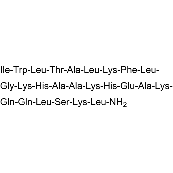
Plantaricin A 是一种抗菌肽,可从植物乳杆菌 (Lactobacillus plantarum) 中提取。Plantaricin A 与环丙沙星具有协同作用。Plantaricin A 表现出抗菌活性。Plantaricin A 可以提高金黄色葡萄球菌的膜电位和细胞内活性氧 (ROS) 水平。Plantaricin A 通过与外排泵结合并改变 MepA、NorA 和 LmrS 的结构,从而抑制外排泵的功能。Plantaricin A 能显著缓解炎症,促进伤口愈合。Plantaricin A 对癌变大鼠垂体细胞具有通透化作用[1][2]。
Patent Blue V calcium salt 是一种用于组织染色和淋巴示踪的三芳基甲烷染料,主要通过局部注射或滴眼应用。Patent Blue V calcium salt 对特定组织 (如角膜内皮、淋巴系统) 具有亲和力,通过吸附或结合使目标结构着色,辅助手术操作。Patent Blue V calcium salt 主要应用于眼科手术 (如 Descemet 膜内皮角膜移植术的移植物染色)、肿瘤前哨淋巴结活检的淋巴引流定位。
专利蓝VF
Acid blue 1 (Patent Blue V) 是一种新型生物染料,可用于视网膜切除术的眼内染料。视网膜切除术是指去除半透明内限制膜 (ILM)。在玻璃体视网膜手术中应用合适染料,才能达到完全去除的目的。Acid blue 1 可被用于染色视网膜膜前结构。光谱分析显示,Acid blue 1 在 450 nm以下和 600 nm 以上都有很强的吸收,表现为蓝绿色。Acid blue 1 还被用作淋巴管造影术的标记物,用于活检手术中染色,以切除淋巴结。
二十二碳六烯酸 (标准品)
Docosahexaenoic acid (Standard) 是 Docosahexaenoic acid 的分析标准品。本产品用于研究及分析应用。Docosahexaenoic Acid (DHA) is an omega-3 fatty acid abundantly present brain and retina. It can be obtained directly from fish oil and maternal milk.
钙黄绿素蓝 (标准品)
Calcein Blue (Standard) 是 Calcein Blue (HY-101887) 的分析标准品。本产品用于研究及分析应用。Calcein Blue 是一种不渗透膜的荧光染料,是一种含有亚氨基二乙酸结构的香豆素衍生物。Calcein Blue 也是一种金属荧光变色指示剂。
专利蓝VF
Acid blue 1 (Patent Blue V) 是一种新型生物染料,可用于视网膜切除术的眼内染料。视网膜切除术是指去除半透明内限制膜 (ILM)。在玻璃体视网膜手术中应用合适染料,才能达到完全去除的目的。Acid blue 1 可被用于染色视网膜膜前结构。光谱分析显示,Acid blue 1 在 450 nm以下和 600 nm 以上都有很强的吸收,表现为蓝绿色。Acid blue 1 还被用作淋巴管造影术的标记物,用于活检手术中染色,以切除淋巴结。
Cardiotoxin Analog (CTX) IV (6-12) 是一种特异性靶向带负电荷磷脂膜 (如磷脂酰丝氨酸、磷脂酰肌醇) 的膜活性肽段,能够从台湾眼镜蛇的毒液中分离得到。Cardiotoxin Analog (CTX) IV (6-12) 是一种蛇毒心脏毒素,通过疏水相互作用和静电吸引结合细胞膜、嵌入脂质双层,从而破坏膜结构稳定性。Cardiotoxin Analog (CTX) IV (6-12) 可诱导膜脂紊乱和细胞裂解,展现出溶血和细胞毒性。
Cardiotoxin Analog (CTX) IV (6-12) TFA 是一种特异性靶向带负电荷磷脂膜 (如磷脂酰丝氨酸、磷脂酰肌醇) 的膜活性肽段,能够从台湾眼镜蛇的毒液中分离得到。Cardiotoxin Analog (CTX) IV (6-12) TFA 是一种蛇毒心脏毒素,通过疏水相互作用和静电吸引结合细胞膜、嵌入脂质双层,从而破坏膜结构稳定性。Cardiotoxin Analog (CTX) IV (6-12) TFA 可诱导膜脂紊乱和细胞裂解,展现出溶血和细胞毒性。
短杆菌肽 S
Gramicidin S (Gramicidin soviet) 是一种选择性靶向细菌细胞膜的阳离子环肽抗生素,具有抗癌活性。Gramicidin S 还破坏膜完整性和干扰膜蛋白功能发挥抗菌活性。Gramicidin S 通过疏水氨基酸残基插入磷脂双层,特异性结合负电荷膜脂并扰乱膜结构,进而抑制细胞分裂和细胞壁合成,最终引发细菌死亡。Gramicidin S 还抑制离子通道,对 Na+/K+-ATPase,烟草叶质膜 Mg2+/K+-ATPase,大鼠心脏质膜 Ca2+-ATPase 的 IC50 分别为 41 μM,24 μM,3 μM。
Plantaricin A 是一种抗菌肽,可从植物乳杆菌 (Lactobacillus plantarum) 中提取。Plantaricin A 与环丙沙星具有协同作用。Plantaricin A 表现出抗菌活性。Plantaricin A 可以提高金黄色葡萄球菌的膜电位和细胞内活性氧 (ROS) 水平。Plantaricin A 通过与外排泵结合并改变 MepA、NorA 和 LmrS 的结构,从而抑制外排泵的功能。Plantaricin A 能显著缓解炎症,促进伤口愈合。Plantaricin A 对癌变大鼠垂体细胞具有通透化作用[1][2]。
Cardiotoxin Analog (CTX) IV (6-12) 是一种特异性靶向带负电荷磷脂膜 (如磷脂酰丝氨酸、磷脂酰肌醇) 的膜活性肽段,能够从台湾眼镜蛇的毒液中分离得到。Cardiotoxin Analog (CTX) IV (6-12) 是一种蛇毒心脏毒素,通过疏水相互作用和静电吸引结合细胞膜、嵌入脂质双层,从而破坏膜结构稳定性。Cardiotoxin Analog (CTX) IV (6-12) 可诱导膜脂紊乱和细胞裂解,展现出溶血和细胞毒性。
Cardiotoxin Analog (CTX) IV (6-12) TFA 是一种特异性靶向带负电荷磷脂膜 (如磷脂酰丝氨酸、磷脂酰肌醇) 的膜活性肽段,能够从台湾眼镜蛇的毒液中分离得到。Cardiotoxin Analog (CTX) IV (6-12) TFA 是一种蛇毒心脏毒素,通过疏水相互作用和静电吸引结合细胞膜、嵌入脂质双层,从而破坏膜结构稳定性。Cardiotoxin Analog (CTX) IV (6-12) TFA 可诱导膜脂紊乱和细胞裂解,展现出溶血和细胞毒性。
二十二碳六烯酸 (标准品)
Docosahexaenoic acid (Standard) 是 Docosahexaenoic acid 的分析标准品。本产品用于研究及分析应用。Docosahexaenoic Acid (DHA) is an omega-3 fatty acid abundantly present brain and retina. It can be obtained directly from fish oil and maternal milk.
Western blot analysis of extracts from THP-1(lane 2(20μg), Jurkat (lane 3(20μg) and NIH3T3(lane 4(20μg) using FOXO1A (HY-P80132) Rabbit mAb. Proteins were transferred
to a PVDF membrane and blocked with 5% non-fat milk in TBST for 2 hour at room temperature. The primary antibody (1/1000) and Loading control antibody (Beta Actin, HY-P80438, 1/10000) was
used in 5% non-fat milk in TBST at 4°C overnight. Goat Anti-Mouse/Rabbit IgG-HRP Secondary Antibody (1/10000) was used for 1 hour at room temperature.
Western blot analysis of extracts from THP-1(lane 2(20μg), Jurkat (lane 3(20μg) and NIH3T3(lane 4(20μg) using FOXO1A (HY-P80132) Rabbit mAb. Proteins were transferred
to a PVDF membrane and blocked with 5% non-fat milk in TBST for 2 hour at room temperature. The primary antibody (1/1000) and Loading control antibody (Beta Actin, HY-P80438, 1/10000) was
MedchemExpress Validation 03
Western blot analysis of extracts from THP-1(lane 2(20μg), Jurkat (lane 3(20μg) and NIH3T3(lane 4(20μg) using FOXO1A (HY-P80132) Rabbit mAb. Proteins were transferred
MedchemExpress Validation 04
Western blot analysis of extracts from THP-1(lane 2(20μg), Jurkat (lane 3(20μg) and NIH3T3(lane 4(20μg) using FOXO1A (HY-P80132) Rabbit mAb. Proteins were transferred
to a PVDF membrane and blocked with 5% non-fat milk in TBST for 2 hour at room temperature. The primary antibody (1/1000) and Loading control antibody (Beta Actin, HY-P80438, 1/10000) was
used in 5% non-fat milk in TBST at 4°C overnight. Goat Anti-Mouse/Rabbit IgG-HRP Secondary Antibody (1/10000) was used for 1 hour at room temperature.
MedchemExpress Validation
Western blot analysis of extracts from THP-1(lane 2(20μg), Jurkat (lane 3(20μg) and NIH3T3(lane 4(20μg) using FOXO1A (HY-P80132) Rabbit mAb. Proteins were transferred
to a PVDF membrane and blocked with 5% non-fat milk in TBST for 2 hour at room temperature. The primary antibody (1/1000) and Loading control antibody (Beta Actin, HY-P80438, 1/10000) was
used in 5% non-fat milk in TBST at 4°C overnight. Goat Anti-Mouse/Rabbit IgG-HRP Secondary Antibody (1/10000) was used for 1 hour at room temperature.
MedchemExpress Validation
Western blot analysis of extracts from THP-1(lane 2(20μg), Jurkat (lane 3(20μg) and NIH3T3(lane 4(20μg) using FOXO1A (HY-P80132) Rabbit mAb. Proteins were transferred
to a PVDF membrane and blocked with 5% non-fat milk in TBST for 2 hour at room temperature. The primary antibody (1/1000) and Loading control antibody (Beta Actin, HY-P80438, 1/10000) was
used in 5% non-fat milk in TBST at 4°C overnight. Goat Anti-Mouse/Rabbit IgG-HRP Secondary Antibody (1/10000) was used for 1 hour at room temperature.


![[BMIM]Cl](http://hg.y866.cn/biomolecule/lib/file/product_pic/hy-w016758.gif)