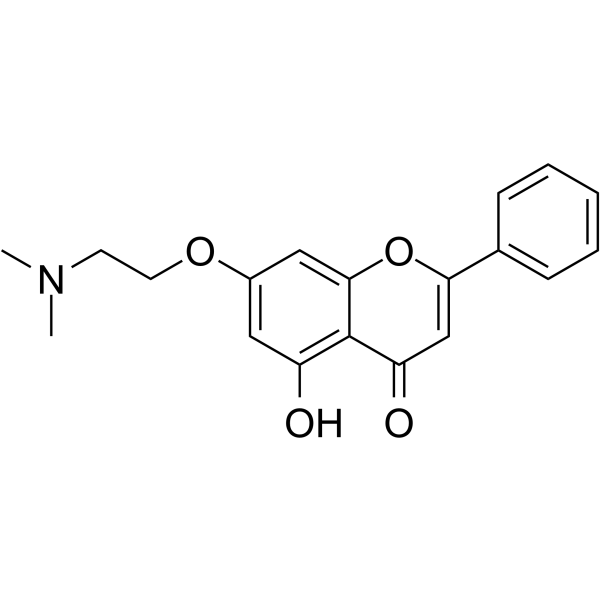
Cannflavin A 可从大麻 (Cannabis sativa L.) 中分离得到。Cannflavin A 具有抗癌、神经保护和抗炎活性。Cannflavin A 可抑制 Aβ1-42 聚集。Cannflavin A 还能抑制犬尿氨酸-3-单加氧酶 (KMO) (IC50 = 29.4 μM)。Cannflavin A 通过 caspase-3 裂解激活细胞凋亡。Cannflavin A 通过抑制促炎酶 (包括 PC12 细胞系中的前列腺素 E2 和细胞色素 c 氧化酶 I 和 II) 发挥抗炎作用。
柴胡皂苷 C (标准品)
Saikosaponin C (Standard) 是 Saikosaponin C 的分析标准品。本产品用于研究及分析应用。Saikosaponin C 是柴胡中的活性成分,在阿尔滋海默症中主要靶作用于 amyloid beta 和 tau 蛋白。Saikosaponin C 抑制 Aβ1-40 和 Aβ1-42 的释放,抑制异常 tau 蛋白的磷酸化,但对 BACE1 的活性和表达无作用。
Cannflavin A (Standard) 是 Cannflavin A 的分析标准品。本产品用于研究及分析应用。Cannflavin A 可从大麻 (Cannabis sativa L.) 中分离得到。Cannflavin A 具有抗癌、神经保护和抗炎活性。Cannflavin A 可抑制 Aβ1-42 聚集。Cannflavin A 还能抑制犬尿氨酸-3-单加氧酶 (KMO) (IC50 = 29.4 μM)。Cannflavin A 通过 caspase-3 裂解激活细胞凋亡。Cannflavin A 通过抑制促炎酶 (包括 PC12 细胞系中的前列腺素 E2 和细胞色素 c 氧化酶 I 和 II) 发挥抗炎作用。
Withanoside V 是一种可透过血脑屏障的睡茄衍生物。Withanoside V 可与人血清白蛋白 (HSA) 的 Sudlow I (结构域 IIA) 强结合,形成稳定复合物并改变白蛋白的二级结构,从而增加螺旋含量、减少 β-折叠和无规卷曲。Withanoside V 可与 Aβ (1-42) 结合,阻断单体间的相互作用及后续聚集。Withanoside V 抑制神经母细胞瘤细胞活力,减少 Aβ (1-42) 诱导凋亡 (apoptosis) 细胞数量,降低 ROS 生成。Withanoside V 可抑制 SARS-CoV-2 Mpro。Withanoside V 可用于阿尔茨海默病、新型冠状病毒病的研究。
Cannflavin A 可从大麻 (Cannabis sativa L.) 中分离得到。Cannflavin A 具有抗癌、神经保护和抗炎活性。Cannflavin A 可抑制 Aβ1-42 聚集。Cannflavin A 还能抑制犬尿氨酸-3-单加氧酶 (KMO) (IC50 = 29.4 μM)。Cannflavin A 通过 caspase-3 裂解激活细胞凋亡。Cannflavin A 通过抑制促炎酶 (包括 PC12 细胞系中的前列腺素 E2 和细胞色素 c 氧化酶 I 和 II) 发挥抗炎作用。
Withanoside V 是一种可透过血脑屏障的睡茄衍生物。Withanoside V 可与人血清白蛋白 (HSA) 的 Sudlow I (结构域 IIA) 强结合,形成稳定复合物并改变白蛋白的二级结构,从而增加螺旋含量、减少 β-折叠和无规卷曲。Withanoside V 可与 Aβ (1-42) 结合,阻断单体间的相互作用及后续聚集。Withanoside V 抑制神经母细胞瘤细胞活力,减少 Aβ (1-42) 诱导凋亡 (apoptosis) 细胞数量,降低 ROS 生成。Withanoside V 可抑制 SARS-CoV-2 Mpro。Withanoside V 可用于阿尔茨海默病、新型冠状病毒病的研究。
柴胡皂苷 C (标准品)
Saikosaponin C (Standard) 是 Saikosaponin C 的分析标准品。本产品用于研究及分析应用。Saikosaponin C 是柴胡中的活性成分,在阿尔滋海默症中主要靶作用于 amyloid beta 和 tau 蛋白。Saikosaponin C 抑制 Aβ1-40 和 Aβ1-42 的释放,抑制异常 tau 蛋白的磷酸化,但对 BACE1 的活性和表达无作用。
Cannflavin A (Standard) 是 Cannflavin A 的分析标准品。本产品用于研究及分析应用。Cannflavin A 可从大麻 (Cannabis sativa L.) 中分离得到。Cannflavin A 具有抗癌、神经保护和抗炎活性。Cannflavin A 可抑制 Aβ1-42 聚集。Cannflavin A 还能抑制犬尿氨酸-3-单加氧酶 (KMO) (IC50 = 29.4 μM)。Cannflavin A 通过 caspase-3 裂解激活细胞凋亡。Cannflavin A 通过抑制促炎酶 (包括 PC12 细胞系中的前列腺素 E2 和细胞色素 c 氧化酶 I 和 II) 发挥抗炎作用。
Western blot analysis of extracts from THP-1(lane 2(20μg), Jurkat (lane 3(20μg) and NIH3T3(lane 4(20μg) using FOXO1A (HY-P80132) Rabbit mAb. Proteins were transferred
to a PVDF membrane and blocked with 5% non-fat milk in TBST for 2 hour at room temperature. The primary antibody (1/1000) and Loading control antibody (Beta Actin, HY-P80438, 1/10000) was
used in 5% non-fat milk in TBST at 4°C overnight. Goat Anti-Mouse/Rabbit IgG-HRP Secondary Antibody (1/10000) was used for 1 hour at room temperature.
Western blot analysis of extracts from THP-1(lane 2(20μg), Jurkat (lane 3(20μg) and NIH3T3(lane 4(20μg) using FOXO1A (HY-P80132) Rabbit mAb. Proteins were transferred
to a PVDF membrane and blocked with 5% non-fat milk in TBST for 2 hour at room temperature. The primary antibody (1/1000) and Loading control antibody (Beta Actin, HY-P80438, 1/10000) was
MedchemExpress Validation 03
Western blot analysis of extracts from THP-1(lane 2(20μg), Jurkat (lane 3(20μg) and NIH3T3(lane 4(20μg) using FOXO1A (HY-P80132) Rabbit mAb. Proteins were transferred
MedchemExpress Validation 04
Western blot analysis of extracts from THP-1(lane 2(20μg), Jurkat (lane 3(20μg) and NIH3T3(lane 4(20μg) using FOXO1A (HY-P80132) Rabbit mAb. Proteins were transferred
to a PVDF membrane and blocked with 5% non-fat milk in TBST for 2 hour at room temperature. The primary antibody (1/1000) and Loading control antibody (Beta Actin, HY-P80438, 1/10000) was
used in 5% non-fat milk in TBST at 4°C overnight. Goat Anti-Mouse/Rabbit IgG-HRP Secondary Antibody (1/10000) was used for 1 hour at room temperature.
MedchemExpress Validation
Western blot analysis of extracts from THP-1(lane 2(20μg), Jurkat (lane 3(20μg) and NIH3T3(lane 4(20μg) using FOXO1A (HY-P80132) Rabbit mAb. Proteins were transferred
to a PVDF membrane and blocked with 5% non-fat milk in TBST for 2 hour at room temperature. The primary antibody (1/1000) and Loading control antibody (Beta Actin, HY-P80438, 1/10000) was
used in 5% non-fat milk in TBST at 4°C overnight. Goat Anti-Mouse/Rabbit IgG-HRP Secondary Antibody (1/10000) was used for 1 hour at room temperature.
MedchemExpress Validation
Western blot analysis of extracts from THP-1(lane 2(20μg), Jurkat (lane 3(20μg) and NIH3T3(lane 4(20μg) using FOXO1A (HY-P80132) Rabbit mAb. Proteins were transferred
to a PVDF membrane and blocked with 5% non-fat milk in TBST for 2 hour at room temperature. The primary antibody (1/1000) and Loading control antibody (Beta Actin, HY-P80438, 1/10000) was
used in 5% non-fat milk in TBST at 4°C overnight. Goat Anti-Mouse/Rabbit IgG-HRP Secondary Antibody (1/10000) was used for 1 hour at room temperature.