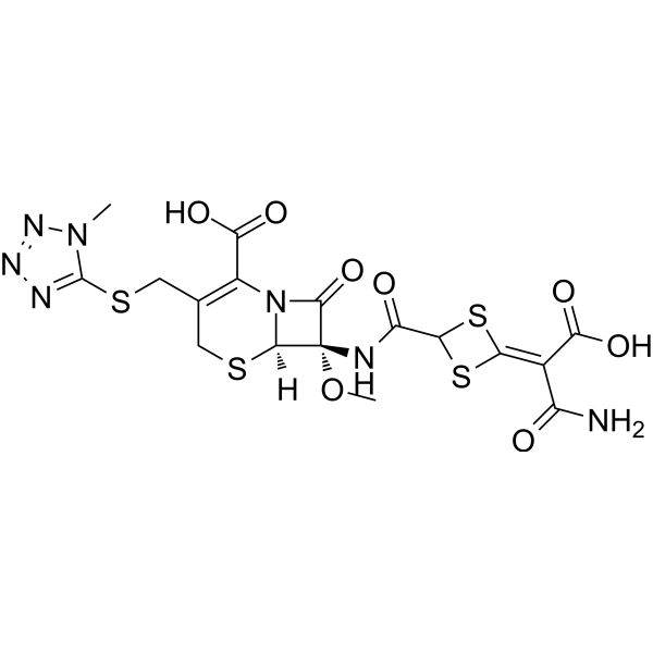
茜素红S指示剂 (C.I.58005)
Alizarin Red S (C.I. 58005) 是一种具有还原活性 (具有醌基结构) 的蒽醌类染料,通过与金属离子 (如锆、钙) 或硼酸形成复合物,发挥钙沉积标记和电化学传感功能。Alizarin Red S 通过可逆氧化还原反应 (用于电化学检测) 和不可逆螯合 (用于骨染色)。Alizarin Red S 主要应用于骨代谢研究 (标记矿化组织) 、糖检测 (硼酸-糖竞争体系) 及金属离子传感 (如锆离子检测),可用于骨质疏松症和代谢疾病研究。
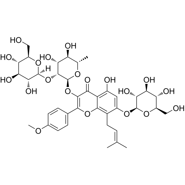
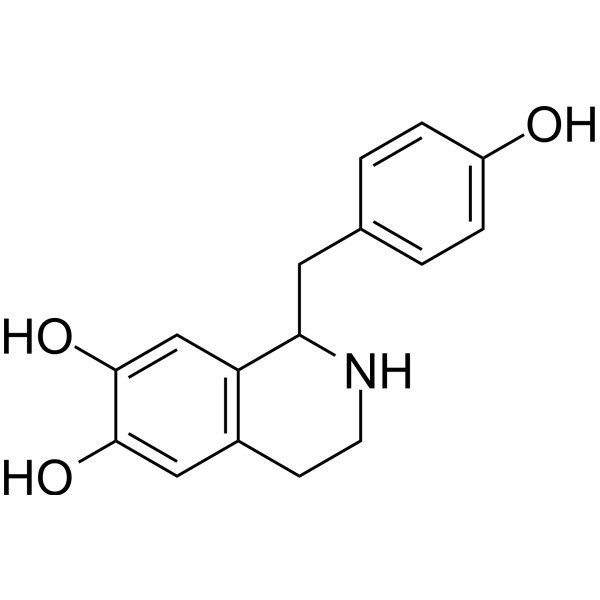
朝藿定A (标准品)
Epimedin A (Standard) 是 Epimedin A (HY-N0257) 的分析标准品。本产品用于研究及分析应用。Epimedin A 是淫羊藿 (Herba Epimedii) 中主要的黄酮类活性成分之一,具有口服活性。Epimedin A 可抑制破骨细胞生成、分化和骨吸收。Epimedin A 具有抗炎活性。Epimedin A 可用于骨质疏松和炎症性疾病的研究。
SM-8849 是一种噻唑衍生物,具有抗关节炎活性。SM-8849特异性靶向并灭活参与迟发型超敏反应 (DTH) 反应的 T 细胞,从而抑制关节炎的核心免疫病理过程,但对抗体产生等体液免疫环节影响很小。SM-8849 在 Type II Collagen (HY-NP003) 诱导的小鼠关节炎模型中显著缓解了临床症状,减轻骨破坏和关节损伤。SM-8849 可用于研究类风湿关节炎等自身免疫性疾病。
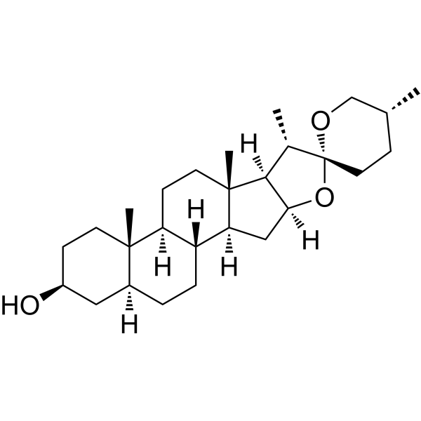
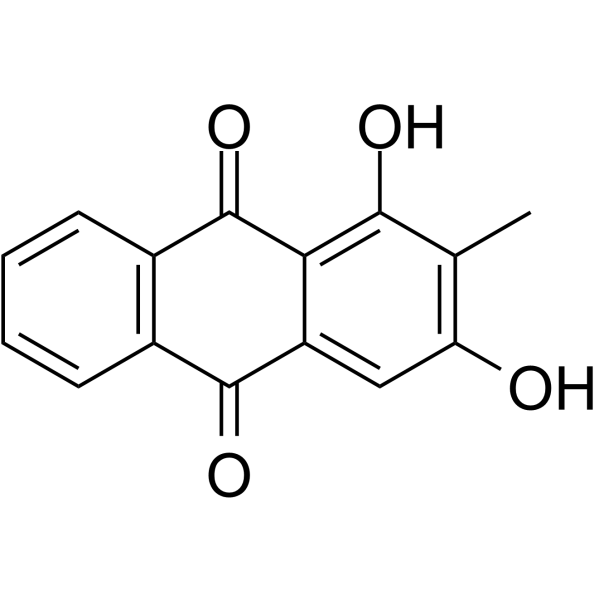
竹节香附素A
Raddeanin A 是一种齐墩果烷型三萜皂苷,具备口服活性。Raddeanin A 可抑制 SRC、mTOR、JNK、VEGFR2、NLRP3 炎症小体、Wnt/β-catenin、Wee1、PI3K/AKT 信号通路、MAPK/ERK 信号通路、AR-FL、AR-Vs,并下调 p-PI3K 和 p-AKT 的表达。Raddeanin A 可抑制破骨细胞形成、骨吸收、溶骨作用、癌细胞侵袭、迁移、增殖、血管生成及上皮-间质转化,同时诱导细胞凋亡 (apoptosis)、细胞周期阻滞、ROS 产生、免疫原性细胞死亡及树突状细胞成熟。Raddeanin A 可改善血视网膜屏障功能、减轻炎症、调控肿瘤微环境,并提升抗 PD-1 抗体的活性。Raddeanin A 可用于乳腺癌相关溶骨症、人骨肉瘤、结直肠癌、胶质母细胞瘤、阿尔茨海默病、胆管癌、黑色素瘤、非小细胞肺癌、去势抵抗性前列腺癌及多发性骨髓瘤的研究。
茜素红S指示剂 (C.I.58005)
Alizarin Red S (C.I. 58005) 是一种具有还原活性 (具有醌基结构) 的蒽醌类染料,通过与金属离子 (如锆、钙) 或硼酸形成复合物,发挥钙沉积标记和电化学传感功能。Alizarin Red S 通过可逆氧化还原反应 (用于电化学检测) 和不可逆螯合 (用于骨染色)。Alizarin Red S 主要应用于骨代谢研究 (标记矿化组织) 、糖检测 (硼酸-糖竞争体系) 及金属离子传感 (如锆离子检测),可用于骨质疏松症和代谢疾病研究。
竹节香附素A
Raddeanin A 是一种齐墩果烷型三萜皂苷,具备口服活性。Raddeanin A 可抑制 SRC、mTOR、JNK、VEGFR2、NLRP3 炎症小体、Wnt/β-catenin、Wee1、PI3K/AKT 信号通路、MAPK/ERK 信号通路、AR-FL、AR-Vs,并下调 p-PI3K 和 p-AKT 的表达。Raddeanin A 可抑制破骨细胞形成、骨吸收、溶骨作用、癌细胞侵袭、迁移、增殖、血管生成及上皮-间质转化,同时诱导细胞凋亡 (apoptosis)、细胞周期阻滞、ROS 产生、免疫原性细胞死亡及树突状细胞成熟。Raddeanin A 可改善血视网膜屏障功能、减轻炎症、调控肿瘤微环境,并提升抗 PD-1 抗体的活性。Raddeanin A 可用于乳腺癌相关溶骨症、人骨肉瘤、结直肠癌、胶质母细胞瘤、阿尔茨海默病、胆管癌、黑色素瘤、非小细胞肺癌、去势抵抗性前列腺癌及多发性骨髓瘤的研究。
朝藿定A (标准品)
Epimedin A (Standard) 是 Epimedin A (HY-N0257) 的分析标准品。本产品用于研究及分析应用。Epimedin A 是淫羊藿 (Herba Epimedii) 中主要的黄酮类活性成分之一,具有口服活性。Epimedin A 可抑制破骨细胞生成、分化和骨吸收。Epimedin A 具有抗炎活性。Epimedin A 可用于骨质疏松和炎症性疾病的研究。
Western blot analysis of extracts from THP-1(lane 2(20μg), Jurkat (lane 3(20μg) and NIH3T3(lane 4(20μg) using FOXO1A (HY-P80132) Rabbit mAb. Proteins were transferred
to a PVDF membrane and blocked with 5% non-fat milk in TBST for 2 hour at room temperature. The primary antibody (1/1000) and Loading control antibody (Beta Actin, HY-P80438, 1/10000) was
used in 5% non-fat milk in TBST at 4°C overnight. Goat Anti-Mouse/Rabbit IgG-HRP Secondary Antibody (1/10000) was used for 1 hour at room temperature.
Western blot analysis of extracts from THP-1(lane 2(20μg), Jurkat (lane 3(20μg) and NIH3T3(lane 4(20μg) using FOXO1A (HY-P80132) Rabbit mAb. Proteins were transferred
to a PVDF membrane and blocked with 5% non-fat milk in TBST for 2 hour at room temperature. The primary antibody (1/1000) and Loading control antibody (Beta Actin, HY-P80438, 1/10000) was
MedchemExpress Validation 03
Western blot analysis of extracts from THP-1(lane 2(20μg), Jurkat (lane 3(20μg) and NIH3T3(lane 4(20μg) using FOXO1A (HY-P80132) Rabbit mAb. Proteins were transferred
MedchemExpress Validation 04
Western blot analysis of extracts from THP-1(lane 2(20μg), Jurkat (lane 3(20μg) and NIH3T3(lane 4(20μg) using FOXO1A (HY-P80132) Rabbit mAb. Proteins were transferred
to a PVDF membrane and blocked with 5% non-fat milk in TBST for 2 hour at room temperature. The primary antibody (1/1000) and Loading control antibody (Beta Actin, HY-P80438, 1/10000) was
used in 5% non-fat milk in TBST at 4°C overnight. Goat Anti-Mouse/Rabbit IgG-HRP Secondary Antibody (1/10000) was used for 1 hour at room temperature.
MedchemExpress Validation
Western blot analysis of extracts from THP-1(lane 2(20μg), Jurkat (lane 3(20μg) and NIH3T3(lane 4(20μg) using FOXO1A (HY-P80132) Rabbit mAb. Proteins were transferred
to a PVDF membrane and blocked with 5% non-fat milk in TBST for 2 hour at room temperature. The primary antibody (1/1000) and Loading control antibody (Beta Actin, HY-P80438, 1/10000) was
used in 5% non-fat milk in TBST at 4°C overnight. Goat Anti-Mouse/Rabbit IgG-HRP Secondary Antibody (1/10000) was used for 1 hour at room temperature.
MedchemExpress Validation
Western blot analysis of extracts from THP-1(lane 2(20μg), Jurkat (lane 3(20μg) and NIH3T3(lane 4(20μg) using FOXO1A (HY-P80132) Rabbit mAb. Proteins were transferred
to a PVDF membrane and blocked with 5% non-fat milk in TBST for 2 hour at room temperature. The primary antibody (1/1000) and Loading control antibody (Beta Actin, HY-P80438, 1/10000) was
used in 5% non-fat milk in TBST at 4°C overnight. Goat Anti-Mouse/Rabbit IgG-HRP Secondary Antibody (1/10000) was used for 1 hour at room temperature.