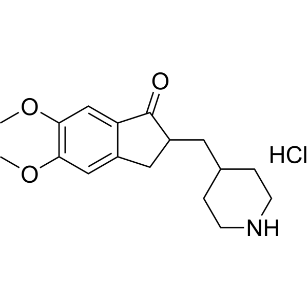
乙酰胆碱酯酶
Acetylcholinesterase, Fly head (ACHE; EC 3.1.1.7), 即乙酰胆碱酯酶,是一种胆碱能酶,主要存在于神经肌肉接头和胆碱能类型的化学突触中,常用于生化研究。Acetylcholinesterase, Fly head 可以催化乙酰胆碱和其他一些充当神经递质的胆碱酯分解或水解为乙酸和胆碱。Acetylcholinesterase, Fly head 主要作用是终止突触之间的神经元传递和信号传导,以防止 ACh 扩散和附近受体的激活。
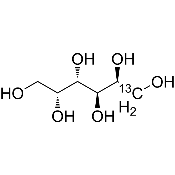
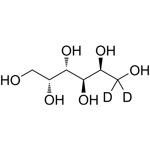
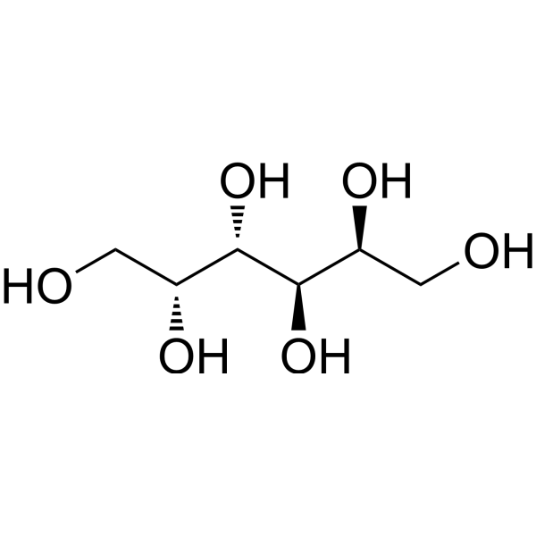
Acetoacetyl CoA 是甲羟戊酸途径中 HMG-CoA 的前体。Acetoacetyl-CoA thiolase 催化两个 acetyl-CoA 分子形成 Acetoacetyl CoA。Acetoacetyl CoA 对胆固醇生物合成至关重要。Acetoacetyl CoA 也是脂肪酸的生物分解和合成的中间体。
重组胶原蛋白酶H
Collagenase H Recombinant 够消化和分解细胞外基质 (ECM) 中的胶原蛋白 (尤其是胶原蛋白 III)。Collagenase H Recombinant 在大鼠胰岛的分离和功能中起着重要作用。Collagenase H Recombinant 可用于组织工程和细胞培养。
核糖核酸酶A
RNase A (Bovine pancreatic RNase) 是一种广泛用于 DNA 纯化的核酸内切酶 (Endonuclease),它能特异性水解 RNA 中的胞嘧啶或尿嘧啶残基。RNase A 可降解 RNA/DNA 双链体中的 RNA。RNase A 催化单链 RNA 的 3',5'-磷酸二酯键断裂。生物体内的 RNase A 家族成员与多种生理和病理过程密切相关,包括细胞生长发育、增殖、分化和迁移。RNase A 活性或表达水平的失调与胰腺癌、卵巢癌、膀胱癌和甲状腺癌密切相关。RNase A 具有杀伤肿瘤细胞的能力。
核糖核酸酶 A, 重组
RNase A (Bovine pancreatic RNase) 是一种广泛用于 DNA 纯化的核酸内切酶 (Endonuclease),它能特异性水解 RNA 中的胞嘧啶或尿嘧啶残基。RNase A 可降解 RNA/DNA 双链体中的 RNA。RNase A 催化单链 RNA 的 3',5'-磷酸二酯键断裂。生物体内的 RNase A 家族成员与多种生理和病理过程密切相关,包括细胞生长发育、增殖、分化和迁移。RNase A 活性或表达水平的失调与胰腺癌、卵巢癌、膀胱癌和甲状腺癌密切相关。RNase A 具有杀伤肿瘤细胞的能力。RNase A, Recombinant (Ribonuclease A, Recombinant) 是重组的 RNase A。
重组核糖核酸酶A (无动物源)
RNase A (Bovine pancreatic RNase) 是一种广泛用于 DNA 纯化的核酸内切酶 (Endonuclease),它能特异性水解 RNA 中的胞嘧啶或尿嘧啶残基。RNase A 可降解 RNA/DNA 双链体中的 RNA。RNase A 催化单链 RNA 的 3',5'-磷酸二酯键断裂。生物体内的 RNase A 家族成员与多种生理和病理过程密切相关,包括细胞生长发育、增殖、分化和迁移。RNase A 活性或表达水平的失调与胰腺癌、卵巢癌、膀胱癌和甲状腺癌密切相关。RNase A 具有杀伤肿瘤细胞的能力。RNase A, Recombinant (animal free) 是重组 RNase A,无动物源性成分。
核糖核酸酶 A (不含 DNase 和蛋白酶), 重组
RNase A (Bovine pancreatic RNase) 是一种广泛用于 DNA 纯化的核酸内切酶 (Endonuclease),它能特异性水解 RNA 中的胞嘧啶或尿嘧啶残基。RNase A 可降解 RNA/DNA 双链体中的 RNA。RNase A 催化单链 RNA 的 3',5'-磷酸二酯键断裂。生物体内的 RNase A 家族成员与多种生理和病理过程密切相关,包括细胞生长发育、增殖、分化和迁移。RNase A 活性或表达水平的失调与胰腺癌、卵巢癌、膀胱癌和甲状腺癌密切相关。RNase A 具有杀伤肿瘤细胞的能力。RNase A (DNase & Protease Free), Recombinant 是重组的 RNase A,不含 DNase 和蛋白酶。
RNase A (Bovine pancreatic RNase) 是一种广泛用于 DNA 纯化的核酸内切酶 (Endonuclease),它能特异性水解 RNA 中的胞嘧啶或尿嘧啶残基。RNase A 可降解 RNA/DNA 双链体中的 RNA。RNase A 催化单链 RNA 的 3',5'-磷酸二酯键断裂。生物体内的 RNase A 家族成员与多种生理和病理过程密切相关,包括细胞生长发育、增殖、分化和迁移。RNase A 活性或表达水平的失调与胰腺癌、卵巢癌、膀胱癌和甲状腺癌密切相关。RNase A 具有杀伤肿瘤细胞的能力。RNase A, Bovine Pancreas (DNase & Protease Free) 是牛胰腺来源 RNase A,不含 DNase 和蛋白酶。
Acetoacetyl CoA 是甲羟戊酸途径中 HMG-CoA 的前体。Acetoacetyl-CoA thiolase 催化两个 acetyl-CoA 分子形成 Acetoacetyl CoA。Acetoacetyl CoA 对胆固醇生物合成至关重要。Acetoacetyl CoA 也是脂肪酸的生物分解和合成的中间体。
Acetoacetyl CoA 是甲羟戊酸途径中 HMG-CoA 的前体。Acetoacetyl-CoA thiolase 催化两个 acetyl-CoA 分子形成 Acetoacetyl CoA。Acetoacetyl CoA 对胆固醇生物合成至关重要。Acetoacetyl CoA 也是脂肪酸的生物分解和合成的中间体。
Western blot analysis of extracts from THP-1(lane 2(20μg), Jurkat (lane 3(20μg) and NIH3T3(lane 4(20μg) using FOXO1A (HY-P80132) Rabbit mAb. Proteins were transferred
to a PVDF membrane and blocked with 5% non-fat milk in TBST for 2 hour at room temperature. The primary antibody (1/1000) and Loading control antibody (Beta Actin, HY-P80438, 1/10000) was
used in 5% non-fat milk in TBST at 4°C overnight. Goat Anti-Mouse/Rabbit IgG-HRP Secondary Antibody (1/10000) was used for 1 hour at room temperature.
Western blot analysis of extracts from THP-1(lane 2(20μg), Jurkat (lane 3(20μg) and NIH3T3(lane 4(20μg) using FOXO1A (HY-P80132) Rabbit mAb. Proteins were transferred
to a PVDF membrane and blocked with 5% non-fat milk in TBST for 2 hour at room temperature. The primary antibody (1/1000) and Loading control antibody (Beta Actin, HY-P80438, 1/10000) was
MedchemExpress Validation 03
Western blot analysis of extracts from THP-1(lane 2(20μg), Jurkat (lane 3(20μg) and NIH3T3(lane 4(20μg) using FOXO1A (HY-P80132) Rabbit mAb. Proteins were transferred
MedchemExpress Validation 04
Western blot analysis of extracts from THP-1(lane 2(20μg), Jurkat (lane 3(20μg) and NIH3T3(lane 4(20μg) using FOXO1A (HY-P80132) Rabbit mAb. Proteins were transferred
to a PVDF membrane and blocked with 5% non-fat milk in TBST for 2 hour at room temperature. The primary antibody (1/1000) and Loading control antibody (Beta Actin, HY-P80438, 1/10000) was
used in 5% non-fat milk in TBST at 4°C overnight. Goat Anti-Mouse/Rabbit IgG-HRP Secondary Antibody (1/10000) was used for 1 hour at room temperature.
MedchemExpress Validation
Western blot analysis of extracts from THP-1(lane 2(20μg), Jurkat (lane 3(20μg) and NIH3T3(lane 4(20μg) using FOXO1A (HY-P80132) Rabbit mAb. Proteins were transferred
to a PVDF membrane and blocked with 5% non-fat milk in TBST for 2 hour at room temperature. The primary antibody (1/1000) and Loading control antibody (Beta Actin, HY-P80438, 1/10000) was
used in 5% non-fat milk in TBST at 4°C overnight. Goat Anti-Mouse/Rabbit IgG-HRP Secondary Antibody (1/10000) was used for 1 hour at room temperature.
MedchemExpress Validation
Western blot analysis of extracts from THP-1(lane 2(20μg), Jurkat (lane 3(20μg) and NIH3T3(lane 4(20μg) using FOXO1A (HY-P80132) Rabbit mAb. Proteins were transferred
to a PVDF membrane and blocked with 5% non-fat milk in TBST for 2 hour at room temperature. The primary antibody (1/1000) and Loading control antibody (Beta Actin, HY-P80438, 1/10000) was
used in 5% non-fat milk in TBST at 4°C overnight. Goat Anti-Mouse/Rabbit IgG-HRP Secondary Antibody (1/10000) was used for 1 hour at room temperature.