Human ACKR3 mRNA 能编码人类非典型趋化因子受体 3(ACKR3)蛋白,该蛋白属于 G 蛋白偶联受体家族的一员。ACKR3 是一种典型的趋化因子受体,它通过高亲和力的趋化因子结合来控制趋化因子的水平和定位,这种结合与经典的配体驱动信号转导级联反应无关,而是导致趋化因子的封闭、降解或跨细胞转运。
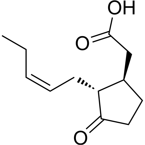
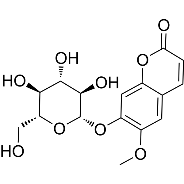
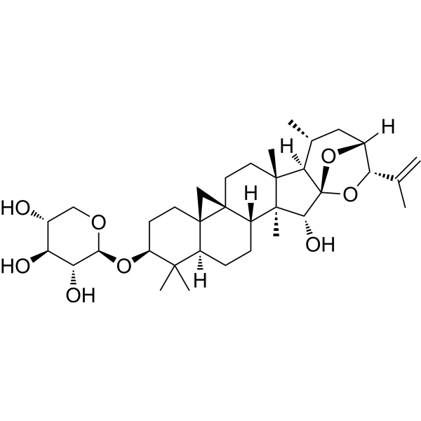
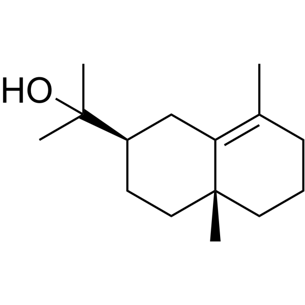
麦冬皂苷D
Ophiopogonin D 是可以从麦冬 (Ophiopogon japonicus) 的块茎中分离的,是一种罕见的天然存在的 C29 甾体糖苷。Ophiopogonin D 是 CYP2J3 诱导剂,其通过增加人脐静脉内皮细胞 (HUVECs) 中 CYP2J2/EETs 和 PPARα 的表达,显着抑制 Ang II 诱导的 NF-κB 核转位,IκBα 下调,细胞内 Ca2+ 过载和促炎细胞因子的激活。Ophiopogonin D 能抑制 RAW264.7 细胞的破骨细胞分化。Ophiopogonin D 作为抗氧化剂,在过氧化氢 (H2O2) 诱导的内皮损伤中具有保护作用。Ophiopogonin D 能阻断 ERK 信号级联。Ophiopogonin D 可缓解高脂饮食引起的代谢综合征,并改变小鼠肠道菌群的结构。Ophiopogonin D 已被用于炎症、代谢和心血管疾病。
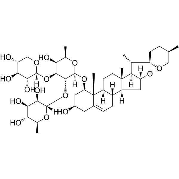
麦冬皂苷D (标准品)
Ophiopogonin D (Standard) 是 Ophiopogonin D 的分析标准品。本产品用于研究及分析应用。Ophiopogonin D 是可以从麦冬 (Ophiopogon japonicus) 的块茎中分离的,是一种罕见的天然存在的 C29 甾体糖苷。Ophiopogonin D 是 CYP2J3 诱导剂,其通过增加人脐静脉内皮细胞 (HUVECs) 中 CYP2J2/EETs 和 PPARα 的表达,显着抑制 Ang II 诱导的 NF-κB 核转位,IκBα 下调,细胞内 Ca2+ 过载和促炎细胞因子的激活。Ophiopogonin D 能抑制 RAW264.7 细胞的破骨细胞分化。Ophiopogonin D 作为抗氧化剂,在过氧化氢 (H2O2) 诱导的内皮损伤中具有保护作用。Ophiopogonin D 能阻断 ERK 信号级联。Ophiopogonin D 可缓解高脂饮食引起的代谢综合征,并改变小鼠肠道菌群的结构。Ophiopogonin D 已被用于炎症、代谢和心血管疾病。
麦冬皂苷D
Ophiopogonin D 是可以从麦冬 (Ophiopogon japonicus) 的块茎中分离的,是一种罕见的天然存在的 C29 甾体糖苷。Ophiopogonin D 是 CYP2J3 诱导剂,其通过增加人脐静脉内皮细胞 (HUVECs) 中 CYP2J2/EETs 和 PPARα 的表达,显着抑制 Ang II 诱导的 NF-κB 核转位,IκBα 下调,细胞内 Ca2+ 过载和促炎细胞因子的激活。Ophiopogonin D 能抑制 RAW264.7 细胞的破骨细胞分化。Ophiopogonin D 作为抗氧化剂,在过氧化氢 (H2O2) 诱导的内皮损伤中具有保护作用。Ophiopogonin D 能阻断 ERK 信号级联。Ophiopogonin D 可缓解高脂饮食引起的代谢综合征,并改变小鼠肠道菌群的结构。Ophiopogonin D 已被用于炎症、代谢和心血管疾病。
麦冬皂苷D (标准品)
Ophiopogonin D (Standard) 是 Ophiopogonin D 的分析标准品。本产品用于研究及分析应用。Ophiopogonin D 是可以从麦冬 (Ophiopogon japonicus) 的块茎中分离的,是一种罕见的天然存在的 C29 甾体糖苷。Ophiopogonin D 是 CYP2J3 诱导剂,其通过增加人脐静脉内皮细胞 (HUVECs) 中 CYP2J2/EETs 和 PPARα 的表达,显着抑制 Ang II 诱导的 NF-κB 核转位,IκBα 下调,细胞内 Ca2+ 过载和促炎细胞因子的激活。Ophiopogonin D 能抑制 RAW264.7 细胞的破骨细胞分化。Ophiopogonin D 作为抗氧化剂,在过氧化氢 (H2O2) 诱导的内皮损伤中具有保护作用。Ophiopogonin D 能阻断 ERK 信号级联。Ophiopogonin D 可缓解高脂饮食引起的代谢综合征,并改变小鼠肠道菌群的结构。Ophiopogonin D 已被用于炎症、代谢和心血管疾病。
Human ACKR3 mRNA 能编码人类非典型趋化因子受体 3(ACKR3)蛋白,该蛋白属于 G 蛋白偶联受体家族的一员。ACKR3 是一种典型的趋化因子受体,它通过高亲和力的趋化因子结合来控制趋化因子的水平和定位,这种结合与经典的配体驱动信号转导级联反应无关,而是导致趋化因子的封闭、降解或跨细胞转运。
Your information is safe with us. * Required Fields.
Western blot analysis of extracts from THP-1(lane 2(20μg), Jurkat (lane 3(20μg) and NIH3T3(lane 4(20μg) using FOXO1A (HY-P80132) Rabbit mAb. Proteins were transferred
to a PVDF membrane and blocked with 5% non-fat milk in TBST for 2 hour at room temperature. The primary antibody (1/1000) and Loading control antibody (Beta Actin, HY-P80438, 1/10000) was
used in 5% non-fat milk in TBST at 4°C overnight. Goat Anti-Mouse/Rabbit IgG-HRP Secondary Antibody (1/10000) was used for 1 hour at room temperature.
Western blot analysis of extracts from THP-1(lane 2(20μg), Jurkat (lane 3(20μg) and NIH3T3(lane 4(20μg) using FOXO1A (HY-P80132) Rabbit mAb. Proteins were transferred
to a PVDF membrane and blocked with 5% non-fat milk in TBST for 2 hour at room temperature. The primary antibody (1/1000) and Loading control antibody (Beta Actin, HY-P80438, 1/10000) was
MedchemExpress Validation 03
Western blot analysis of extracts from THP-1(lane 2(20μg), Jurkat (lane 3(20μg) and NIH3T3(lane 4(20μg) using FOXO1A (HY-P80132) Rabbit mAb. Proteins were transferred
MedchemExpress Validation 04
Western blot analysis of extracts from THP-1(lane 2(20μg), Jurkat (lane 3(20μg) and NIH3T3(lane 4(20μg) using FOXO1A (HY-P80132) Rabbit mAb. Proteins were transferred
to a PVDF membrane and blocked with 5% non-fat milk in TBST for 2 hour at room temperature. The primary antibody (1/1000) and Loading control antibody (Beta Actin, HY-P80438, 1/10000) was
used in 5% non-fat milk in TBST at 4°C overnight. Goat Anti-Mouse/Rabbit IgG-HRP Secondary Antibody (1/10000) was used for 1 hour at room temperature.
MedchemExpress Validation
Western blot analysis of extracts from THP-1(lane 2(20μg), Jurkat (lane 3(20μg) and NIH3T3(lane 4(20μg) using FOXO1A (HY-P80132) Rabbit mAb. Proteins were transferred
to a PVDF membrane and blocked with 5% non-fat milk in TBST for 2 hour at room temperature. The primary antibody (1/1000) and Loading control antibody (Beta Actin, HY-P80438, 1/10000) was
used in 5% non-fat milk in TBST at 4°C overnight. Goat Anti-Mouse/Rabbit IgG-HRP Secondary Antibody (1/10000) was used for 1 hour at room temperature.
MedchemExpress Validation
Western blot analysis of extracts from THP-1(lane 2(20μg), Jurkat (lane 3(20μg) and NIH3T3(lane 4(20μg) using FOXO1A (HY-P80132) Rabbit mAb. Proteins were transferred
to a PVDF membrane and blocked with 5% non-fat milk in TBST for 2 hour at room temperature. The primary antibody (1/1000) and Loading control antibody (Beta Actin, HY-P80438, 1/10000) was
used in 5% non-fat milk in TBST at 4°C overnight. Goat Anti-Mouse/Rabbit IgG-HRP Secondary Antibody (1/10000) was used for 1 hour at room temperature.