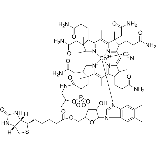
茜素红S指示剂 (C.I.58005)
Alizarin Red S (C.I. 58005) 是一种具有还原活性 (具有醌基结构) 的蒽醌类染料,通过与金属离子 (如锆、钙) 或硼酸形成复合物,发挥钙沉积标记和电化学传感功能。Alizarin Red S 通过可逆氧化还原反应 (用于电化学检测) 和不可逆螯合 (用于骨染色)。Alizarin Red S 主要应用于骨代谢研究 (标记矿化组织) 、糖检测 (硼酸-糖竞争体系) 及金属离子传感 (如锆离子检测),可用于骨质疏松症和代谢疾病研究。
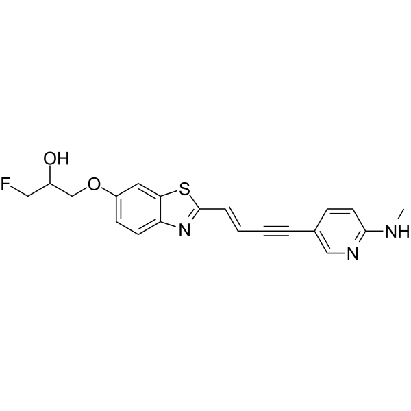
代谢组学是对生物系统中的小分子代谢物进行系统表征的学科,已成为植物科学、微生物生物技术和生物医学研究领域基础研究和转化应用中不可或缺的分析平台。作为多组学整合的重要组成部分,该学科可解读在基因组、转录组和蛋白质组调控下游运行的错综复杂的分子网络,从而捕捉最接近生物体功能的动态生化表型。代谢组由分子量通常低于 1500 Da 的内源化合物组成,是细胞过程和环境相互作用的功能读数,代谢网络的扰动往往与疾病的发病机制有关。这些独特的属性促使代谢组学在药理学研究中发挥着举足轻重的作用,尤其是在靶点解构、药效学评估和病理过程的机理阐明方面。
茜素红S指示剂 (C.I.58005)
Alizarin Red S (C.I. 58005) 是一种具有还原活性 (具有醌基结构) 的蒽醌类染料,通过与金属离子 (如锆、钙) 或硼酸形成复合物,发挥钙沉积标记和电化学传感功能。Alizarin Red S 通过可逆氧化还原反应 (用于电化学检测) 和不可逆螯合 (用于骨染色)。Alizarin Red S 主要应用于骨代谢研究 (标记矿化组织) 、糖检测 (硼酸-糖竞争体系) 及金属离子传感 (如锆离子检测),可用于骨质疏松症和代谢疾病研究。
Western blot analysis of extracts from THP-1(lane 2(20μg), Jurkat (lane 3(20μg) and NIH3T3(lane 4(20μg) using FOXO1A (HY-P80132) Rabbit mAb. Proteins were transferred
to a PVDF membrane and blocked with 5% non-fat milk in TBST for 2 hour at room temperature. The primary antibody (1/1000) and Loading control antibody (Beta Actin, HY-P80438, 1/10000) was
used in 5% non-fat milk in TBST at 4°C overnight. Goat Anti-Mouse/Rabbit IgG-HRP Secondary Antibody (1/10000) was used for 1 hour at room temperature.
Western blot analysis of extracts from THP-1(lane 2(20μg), Jurkat (lane 3(20μg) and NIH3T3(lane 4(20μg) using FOXO1A (HY-P80132) Rabbit mAb. Proteins were transferred
to a PVDF membrane and blocked with 5% non-fat milk in TBST for 2 hour at room temperature. The primary antibody (1/1000) and Loading control antibody (Beta Actin, HY-P80438, 1/10000) was
MedchemExpress Validation 03
Western blot analysis of extracts from THP-1(lane 2(20μg), Jurkat (lane 3(20μg) and NIH3T3(lane 4(20μg) using FOXO1A (HY-P80132) Rabbit mAb. Proteins were transferred
MedchemExpress Validation 04
Western blot analysis of extracts from THP-1(lane 2(20μg), Jurkat (lane 3(20μg) and NIH3T3(lane 4(20μg) using FOXO1A (HY-P80132) Rabbit mAb. Proteins were transferred
to a PVDF membrane and blocked with 5% non-fat milk in TBST for 2 hour at room temperature. The primary antibody (1/1000) and Loading control antibody (Beta Actin, HY-P80438, 1/10000) was
used in 5% non-fat milk in TBST at 4°C overnight. Goat Anti-Mouse/Rabbit IgG-HRP Secondary Antibody (1/10000) was used for 1 hour at room temperature.
MedchemExpress Validation
Western blot analysis of extracts from THP-1(lane 2(20μg), Jurkat (lane 3(20μg) and NIH3T3(lane 4(20μg) using FOXO1A (HY-P80132) Rabbit mAb. Proteins were transferred
to a PVDF membrane and blocked with 5% non-fat milk in TBST for 2 hour at room temperature. The primary antibody (1/1000) and Loading control antibody (Beta Actin, HY-P80438, 1/10000) was
used in 5% non-fat milk in TBST at 4°C overnight. Goat Anti-Mouse/Rabbit IgG-HRP Secondary Antibody (1/10000) was used for 1 hour at room temperature.
MedchemExpress Validation
Western blot analysis of extracts from THP-1(lane 2(20μg), Jurkat (lane 3(20μg) and NIH3T3(lane 4(20μg) using FOXO1A (HY-P80132) Rabbit mAb. Proteins were transferred
to a PVDF membrane and blocked with 5% non-fat milk in TBST for 2 hour at room temperature. The primary antibody (1/1000) and Loading control antibody (Beta Actin, HY-P80438, 1/10000) was
used in 5% non-fat milk in TBST at 4°C overnight. Goat Anti-Mouse/Rabbit IgG-HRP Secondary Antibody (1/10000) was used for 1 hour at room temperature.