MitoSOX Red 是特异靶向线粒体的活细胞荧光探针,具有细胞膜渗透性。MitoSOX Red 进入线粒体后会被超氧化物氧化,而不会被其他 ROS 或 RNS 生成系统氧化。被氧化的 MitoSOX Red 随后与线粒体/细胞核内的核酸结合,产生强红色荧光。MitoSOX Red 可以作为荧光指示剂,特异性检测超氧化物。另外,超氧化物歧化酶 (Superoxide dismutase,SOD) 能够预防 MitoSOX Red 氧化。 激发/发射波长:510/580 nm。
1,9-Dimethylmethylene blue 是一种作为亚甲蓝衍生的光敏剂、病毒灭活剂和血红蛋白氧化剂。1,9-Dimethylmethylene blue 被激活时能产生包括单线态氧在内的活性氧,可用作异染性染料。1,9-Dimethylmethylene blue 以单体或二聚体形式被激活时,分别通过非单线态氧活性氧或单线态氧介导的途径诱导 R17 噬菌体和水疱性口炎病毒光灭活并氧化血红蛋白,其中单体形式因具有更高的核酸亲和力,能在特定条件下实现病毒灭活而不形成高铁血红蛋白。1,9-Dimethylmethylene blue 可与糖胺聚糖等物质结合产生颜色变化,虽易受尿液中非糖胺聚糖成分干扰,但仍可用于糖胺聚糖定量的分光光度分析。凭借这些独特的光化学和结合特性,1,9-Dimethylmethylene blue 被广泛应用于病毒感染及相关生化分析的研究中。
MitoSOX Red (solution) 是特异靶向线粒体的活细胞荧光探针,具有细胞膜渗透性。MitoSOX Red 进入线粒体后会被超氧化物氧化,而不会被其他 ROS 或 RNS 生成系统氧化。被氧化的 MitoSOX Red 随后与线粒体/细胞核内的核酸结合,产生强红色荧光。MitoSOX Red 可以作为荧光指示剂,特异性检测超氧化物。另外,超氧化物歧化酶 (Superoxide dismutase,SOD) 能够预防 MitoSOX Red 氧化。 激发/发射波长:510/580 nm。 溶剂及浓度:DMSO:1 mM
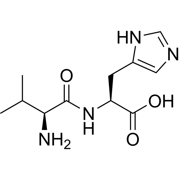
L-肌肽 (标准品)
L-Carnosine (Standard) 是 L-Carnosine 的分析标准品。本产品用于研究及分析应用。L-Carnosine is a dipeptide of the amino acids beta-alanine and histidine and has the potential to suppress many of the biochemical changes that accompany aging.
Tempo (Standard)是 Tempo 的分析标准品。本产品用于研究及分析应用。Tempo 是一氧化氮自由基,也是一种线粒体中 ROS 的选择性清除剂。Tempo 还是一种有机催化剂,可在催化循环中使超氧化物歧化,将伯醇氧化为醛。Tempo 具有诱变和抗氧化作用,可诱导 DNA 链断裂。Tempo 还在小鼠淋巴瘤细胞中发挥细胞毒性和诱变性。
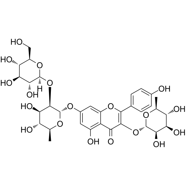
吉奥诺苷D
Jionoside D 是一种具有抗氧化作用的羟基肉桂酸酯。Jionoside D 具有清除胞内活性氧 (ROS) 和 DPPH 自由基的活性,以及抑制脂质过氧化的活性。Jionoside D 可降低 H2O2 诱导的 V79-4 细胞凋亡 (apoptosis)。Jionoside D 可提高细胞抗氧化酶、SOD 和过氧化氢酶的活性。
5-羟基芦荟苷A
5-Hydroxyaloin A 是一种多酚类抗氧化剂。5-Hydroxyaloin A 可在脂肪酶的活性位点和超氧化物歧化酶 (SOD) 的活性位点形成结合能较低的氢键相互作用。5-Hydroxyaloin A 可抑制半胱氨酸亚铁诱导的微粒体脂质过氧化,减少丙二醛的生成。5-Hydroxyaloin A 可用于肥胖症的研究。
MitoSOX Red 是特异靶向线粒体的活细胞荧光探针,具有细胞膜渗透性。MitoSOX Red 进入线粒体后会被超氧化物氧化,而不会被其他 ROS 或 RNS 生成系统氧化。被氧化的 MitoSOX Red 随后与线粒体/细胞核内的核酸结合,产生强红色荧光。MitoSOX Red 可以作为荧光指示剂,特异性检测超氧化物。另外,超氧化物歧化酶 (Superoxide dismutase,SOD) 能够预防 MitoSOX Red 氧化。 激发/发射波长:510/580 nm。
MitoSOX Red (solution) 是特异靶向线粒体的活细胞荧光探针,具有细胞膜渗透性。MitoSOX Red 进入线粒体后会被超氧化物氧化,而不会被其他 ROS 或 RNS 生成系统氧化。被氧化的 MitoSOX Red 随后与线粒体/细胞核内的核酸结合,产生强红色荧光。MitoSOX Red 可以作为荧光指示剂,特异性检测超氧化物。另外,超氧化物歧化酶 (Superoxide dismutase,SOD) 能够预防 MitoSOX Red 氧化。 激发/发射波长:510/580 nm。 溶剂及浓度:DMSO:1 mM
L-肌肽 (标准品)
L-Carnosine (Standard) 是 L-Carnosine 的分析标准品。本产品用于研究及分析应用。L-Carnosine is a dipeptide of the amino acids beta-alanine and histidine and has the potential to suppress many of the biochemical changes that accompany aging.
L-肌肽 (标准品)
L-Carnosine (Standard) 是 L-Carnosine 的分析标准品。本产品用于研究及分析应用。L-Carnosine is a dipeptide of the amino acids beta-alanine and histidine and has the potential to suppress many of the biochemical changes that accompany aging.
吉奥诺苷D
Jionoside D 是一种具有抗氧化作用的羟基肉桂酸酯。Jionoside D 具有清除胞内活性氧 (ROS) 和 DPPH 自由基的活性,以及抑制脂质过氧化的活性。Jionoside D 可降低 H2O2 诱导的 V79-4 细胞凋亡 (apoptosis)。Jionoside D 可提高细胞抗氧化酶、SOD 和过氧化氢酶的活性。
5-羟基芦荟苷A
5-Hydroxyaloin A 是一种多酚类抗氧化剂。5-Hydroxyaloin A 可在脂肪酶的活性位点和超氧化物歧化酶 (SOD) 的活性位点形成结合能较低的氢键相互作用。5-Hydroxyaloin A 可抑制半胱氨酸亚铁诱导的微粒体脂质过氧化,减少丙二醛的生成。5-Hydroxyaloin A 可用于肥胖症的研究。
Western blot analysis of extracts from THP-1(lane 2(20μg), Jurkat (lane 3(20μg) and NIH3T3(lane 4(20μg) using FOXO1A (HY-P80132) Rabbit mAb. Proteins were transferred
to a PVDF membrane and blocked with 5% non-fat milk in TBST for 2 hour at room temperature. The primary antibody (1/1000) and Loading control antibody (Beta Actin, HY-P80438, 1/10000) was
used in 5% non-fat milk in TBST at 4°C overnight. Goat Anti-Mouse/Rabbit IgG-HRP Secondary Antibody (1/10000) was used for 1 hour at room temperature.
Western blot analysis of extracts from THP-1(lane 2(20μg), Jurkat (lane 3(20μg) and NIH3T3(lane 4(20μg) using FOXO1A (HY-P80132) Rabbit mAb. Proteins were transferred
to a PVDF membrane and blocked with 5% non-fat milk in TBST for 2 hour at room temperature. The primary antibody (1/1000) and Loading control antibody (Beta Actin, HY-P80438, 1/10000) was
MedchemExpress Validation 03
Western blot analysis of extracts from THP-1(lane 2(20μg), Jurkat (lane 3(20μg) and NIH3T3(lane 4(20μg) using FOXO1A (HY-P80132) Rabbit mAb. Proteins were transferred
MedchemExpress Validation 04
Western blot analysis of extracts from THP-1(lane 2(20μg), Jurkat (lane 3(20μg) and NIH3T3(lane 4(20μg) using FOXO1A (HY-P80132) Rabbit mAb. Proteins were transferred
to a PVDF membrane and blocked with 5% non-fat milk in TBST for 2 hour at room temperature. The primary antibody (1/1000) and Loading control antibody (Beta Actin, HY-P80438, 1/10000) was
used in 5% non-fat milk in TBST at 4°C overnight. Goat Anti-Mouse/Rabbit IgG-HRP Secondary Antibody (1/10000) was used for 1 hour at room temperature.
MedchemExpress Validation
Western blot analysis of extracts from THP-1(lane 2(20μg), Jurkat (lane 3(20μg) and NIH3T3(lane 4(20μg) using FOXO1A (HY-P80132) Rabbit mAb. Proteins were transferred
to a PVDF membrane and blocked with 5% non-fat milk in TBST for 2 hour at room temperature. The primary antibody (1/1000) and Loading control antibody (Beta Actin, HY-P80438, 1/10000) was
used in 5% non-fat milk in TBST at 4°C overnight. Goat Anti-Mouse/Rabbit IgG-HRP Secondary Antibody (1/10000) was used for 1 hour at room temperature.
MedchemExpress Validation
Western blot analysis of extracts from THP-1(lane 2(20μg), Jurkat (lane 3(20μg) and NIH3T3(lane 4(20μg) using FOXO1A (HY-P80132) Rabbit mAb. Proteins were transferred
to a PVDF membrane and blocked with 5% non-fat milk in TBST for 2 hour at room temperature. The primary antibody (1/1000) and Loading control antibody (Beta Actin, HY-P80438, 1/10000) was
used in 5% non-fat milk in TBST at 4°C overnight. Goat Anti-Mouse/Rabbit IgG-HRP Secondary Antibody (1/10000) was used for 1 hour at room temperature.