脂多糖,来源于大肠杆菌O128:B12
Lipopolysaccharides, from E. coli O128:B12 (LPS, from Escherichia coli (O128:B12)) 是从大肠杆菌 (E. coli O128:B12) 中提取的脂多糖内毒素和 TLR-4 激活剂,是一种 S (smooth) 型 LPS。Lipopolysaccharides, from E. coli O128:B12 具有典型的 3 部分结构:O 抗原、R3 型核心寡糖和脂质 A。Lipopolysaccharides, from E. coli O128:B12 激活免疫细胞的 TLR-4,可用于构建动物新生儿脑部炎症模型,并可能影响新生儿早产。
建议配制浓度 ≥2 mg/mL,充分涡旋震荡 10 分钟以上,必要时辅助超声。由于 LPS 具有吸附特性,分装保存时需使用硅烷化容器或低吸附离心管,使用前充分混匀。
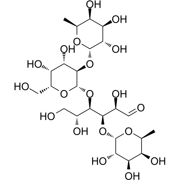
多杀菌素D苷元
Spinosyn D aglycone (A-83543D aglycone)是杀虫剂 Spinosyn D (HY-125326) 的一种苷元形式。与 spinosyn D 不同,spinosyn D 苷元对 H. virescens(烟芽虫)新生幼虫无害(LC50=>64 ppm)。
脂多糖,来源于大肠杆菌O128:B12
Lipopolysaccharides, from E. coli O128:B12 (LPS, from Escherichia coli (O128:B12)) 是从大肠杆菌 (E. coli O128:B12) 中提取的脂多糖内毒素和 TLR-4 激活剂,是一种 S (smooth) 型 LPS。Lipopolysaccharides, from E. coli O128:B12 具有典型的 3 部分结构:O 抗原、R3 型核心寡糖和脂质 A。Lipopolysaccharides, from E. coli O128:B12 激活免疫细胞的 TLR-4,可用于构建动物新生儿脑部炎症模型,并可能影响新生儿早产。
建议配制浓度 ≥2 mg/mL,充分涡旋震荡 10 分钟以上,必要时辅助超声。由于 LPS 具有吸附特性,分装保存时需使用硅烷化容器或低吸附离心管,使用前充分混匀。
Western blot analysis of extracts from THP-1(lane 2(20μg), Jurkat (lane 3(20μg) and NIH3T3(lane 4(20μg) using FOXO1A (HY-P80132) Rabbit mAb. Proteins were transferred
to a PVDF membrane and blocked with 5% non-fat milk in TBST for 2 hour at room temperature. The primary antibody (1/1000) and Loading control antibody (Beta Actin, HY-P80438, 1/10000) was
used in 5% non-fat milk in TBST at 4°C overnight. Goat Anti-Mouse/Rabbit IgG-HRP Secondary Antibody (1/10000) was used for 1 hour at room temperature.
Western blot analysis of extracts from THP-1(lane 2(20μg), Jurkat (lane 3(20μg) and NIH3T3(lane 4(20μg) using FOXO1A (HY-P80132) Rabbit mAb. Proteins were transferred
to a PVDF membrane and blocked with 5% non-fat milk in TBST for 2 hour at room temperature. The primary antibody (1/1000) and Loading control antibody (Beta Actin, HY-P80438, 1/10000) was
MedchemExpress Validation 03
Western blot analysis of extracts from THP-1(lane 2(20μg), Jurkat (lane 3(20μg) and NIH3T3(lane 4(20μg) using FOXO1A (HY-P80132) Rabbit mAb. Proteins were transferred
MedchemExpress Validation 04
Western blot analysis of extracts from THP-1(lane 2(20μg), Jurkat (lane 3(20μg) and NIH3T3(lane 4(20μg) using FOXO1A (HY-P80132) Rabbit mAb. Proteins were transferred
to a PVDF membrane and blocked with 5% non-fat milk in TBST for 2 hour at room temperature. The primary antibody (1/1000) and Loading control antibody (Beta Actin, HY-P80438, 1/10000) was
used in 5% non-fat milk in TBST at 4°C overnight. Goat Anti-Mouse/Rabbit IgG-HRP Secondary Antibody (1/10000) was used for 1 hour at room temperature.
MedchemExpress Validation
Western blot analysis of extracts from THP-1(lane 2(20μg), Jurkat (lane 3(20μg) and NIH3T3(lane 4(20μg) using FOXO1A (HY-P80132) Rabbit mAb. Proteins were transferred
to a PVDF membrane and blocked with 5% non-fat milk in TBST for 2 hour at room temperature. The primary antibody (1/1000) and Loading control antibody (Beta Actin, HY-P80438, 1/10000) was
used in 5% non-fat milk in TBST at 4°C overnight. Goat Anti-Mouse/Rabbit IgG-HRP Secondary Antibody (1/10000) was used for 1 hour at room temperature.
MedchemExpress Validation
Western blot analysis of extracts from THP-1(lane 2(20μg), Jurkat (lane 3(20μg) and NIH3T3(lane 4(20μg) using FOXO1A (HY-P80132) Rabbit mAb. Proteins were transferred
to a PVDF membrane and blocked with 5% non-fat milk in TBST for 2 hour at room temperature. The primary antibody (1/1000) and Loading control antibody (Beta Actin, HY-P80438, 1/10000) was
used in 5% non-fat milk in TBST at 4°C overnight. Goat Anti-Mouse/Rabbit IgG-HRP Secondary Antibody (1/10000) was used for 1 hour at room temperature.