血管紧张素Ⅱ
Angiotensin II (Angiotensin II) 是一种血管收缩剂,是肾素/血管紧张素系统的主要生物活性肽。Angiotensin II human 在调节人类血压中起着核心作用,主要通过血管紧张素 II 与 G 蛋白偶联受体 (GPCRs)、血管紧张素II 1型受体 (AT1R) 和血管紧张素II 2型受体 (AT2R) 之间的相互作用来介导。Angiotensin II human 刺激交感神经刺激,增加醛固酮生物合成和肾脏活动。Angiotensin II human 诱导血管平滑肌细胞生长,增加成纤维细胞中 I 型和 III 型胶原的合成,导致血管壁和心肌增厚,并导致纤维化。Angiotensin II human 也诱导细胞凋亡 (apoptosis)。Angiotensin II human 通过 LOX-1 依赖的氧化还原敏感途径诱导内皮细胞毛细血管形成。
Human serum albumin (HSA) 是血浆中含量最高的蛋白质,是影响血浆瘤压的主要因素。Human serum albumin 具有抗氧化、抗凝血、抗炎、抗血小板聚集活性以及胶体渗透作用。Human serum albumin 能阻止 GML 抑制人类 T 细胞的能力,实现对 T 细胞功能的保护。Human serum albumin 也与心血管疾病相关,能部分阻止 LPS (HY-D1056) 诱导的氧化应激以及血管壁中 NF-κB、iNOS 和 过氧亚硝酸根 (ONOO−) 上调的血压降低。 本产品是在微生物表达系统中重组表达的人血清白蛋白。
血管紧张素 II 醋酸盐
Angiotensin II human (Angiotensin II) acetate 是一种血管收缩剂,是肾素/血管紧张素系统的主要生物活性肽。Angiotensin II human acetate 在调节人类血压中起着核心作用,主要通过血管紧张素 II 与 G 蛋白偶联受体 (GPCRs)、血管紧张素II 1型受体 (AT1R) 和血管紧张素II 2型受体 (AT2R) 之间的相互作用来介导。Angiotensin II human acetate 刺激交感神经刺激,增加醛固酮生物合成和肾脏活动。Angiotensin II human acetate 诱导血管平滑肌细胞生长,增加成纤维细胞中 I 型和 III 型胶原的合成,导致血管壁和心肌增厚,并导致纤维化。Angiotensin II human acetate 也诱导细胞凋亡。Angiotensin II human acetate 通过 LOX-1 依赖的氧化还原敏感途径诱导内皮细胞毛细血管形成。
Angiotensin II human (Angiotensin II) TFA 是一种血管收缩剂,是肾素/血管紧张素系统的主要生物活性肽。Angiotensin II human TFA 在调节人类血压中起着核心作用,主要通过血管紧张素 II 与 G 蛋白偶联受体 (GPCRs)、血管紧张素II 1型受体 (AT1R) 和血管紧张素II 2型受体 (AT2R) 之间的相互作用来介导。Angiotensin II human TFA 刺激交感神经刺激,增加醛固酮生物合成和肾脏活动。Angiotensin II human TFA 诱导血管平滑肌细胞生长,增加成纤维细胞中 I 型和 III 型胶原的合成,导致血管壁和心肌增厚,并导致纤维化。Angiotensin II human TFA 也诱导细胞凋亡。Angiotensin II human TFA 通过 LOX-1 依赖的氧化还原敏感途径诱导内皮细胞毛细血管形成。
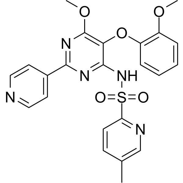
NCX-278 是一种人类碳酸酐酶 (carbonic anhydrase) 抑制剂,对 hCA II 的 Ki 为 13 nM,对 hCA I 的 Ki 为 410 nM,对 hCA IV 的 Ki 为 181 nM,且对 hCA II 的抑制作用相较于 hCA IV 具有选择性。NCX-278 是一种强效的 NO/sGC/cGMP 信号刺激剂,其 EC50 为 2.05 μM。NCX-278 可发挥 NO 介导的血管舒张作用。NCX-278 能够降低正常血压兔的眼内压。NCX-278 可用于开角型青光眼的研究。
Human serum albumin (HSA) 是血浆中含量最高的蛋白质,是影响血浆瘤压的主要因素。Human serum albumin 具有抗氧化、抗凝血、抗炎、抗血小板聚集活性以及胶体渗透作用。Human serum albumin 能阻止 GML 抑制人类 T 细胞的能力,实现对 T 细胞功能的保护。Human serum albumin 也与心血管疾病相关,能部分阻止 LPS (HY-D1056) 诱导的氧化应激以及血管壁中 NF-κB、iNOS 和 过氧亚硝酸根 (ONOO−) 上调的血压降低。 本产品是在微生物表达系统中重组表达的人血清白蛋白。
血管紧张素Ⅱ
Angiotensin II (Angiotensin II) 是一种血管收缩剂,是肾素/血管紧张素系统的主要生物活性肽。Angiotensin II human 在调节人类血压中起着核心作用,主要通过血管紧张素 II 与 G 蛋白偶联受体 (GPCRs)、血管紧张素II 1型受体 (AT1R) 和血管紧张素II 2型受体 (AT2R) 之间的相互作用来介导。Angiotensin II human 刺激交感神经刺激,增加醛固酮生物合成和肾脏活动。Angiotensin II human 诱导血管平滑肌细胞生长,增加成纤维细胞中 I 型和 III 型胶原的合成,导致血管壁和心肌增厚,并导致纤维化。Angiotensin II human 也诱导细胞凋亡 (apoptosis)。Angiotensin II human 通过 LOX-1 依赖的氧化还原敏感途径诱导内皮细胞毛细血管形成。
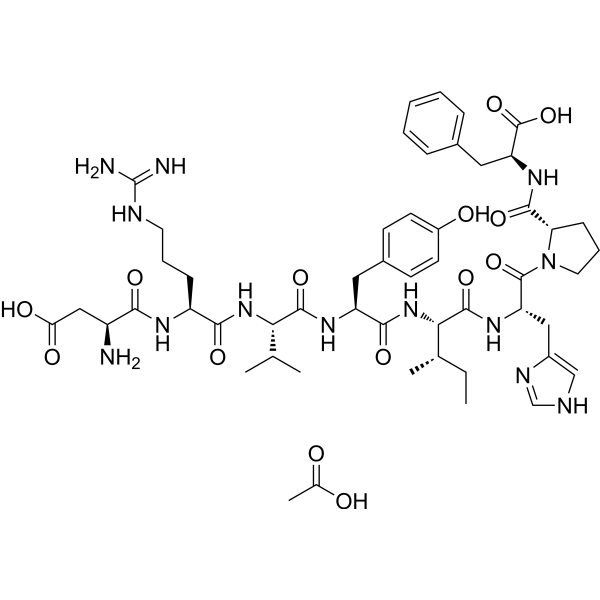
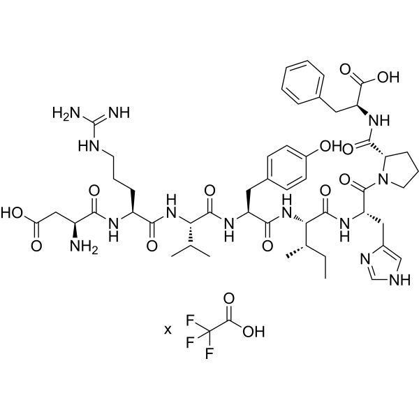
血管紧张素 II 醋酸盐
Angiotensin II human (Angiotensin II) acetate 是一种血管收缩剂,是肾素/血管紧张素系统的主要生物活性肽。Angiotensin II human acetate 在调节人类血压中起着核心作用,主要通过血管紧张素 II 与 G 蛋白偶联受体 (GPCRs)、血管紧张素II 1型受体 (AT1R) 和血管紧张素II 2型受体 (AT2R) 之间的相互作用来介导。Angiotensin II human acetate 刺激交感神经刺激,增加醛固酮生物合成和肾脏活动。Angiotensin II human acetate 诱导血管平滑肌细胞生长,增加成纤维细胞中 I 型和 III 型胶原的合成,导致血管壁和心肌增厚,并导致纤维化。Angiotensin II human acetate 也诱导细胞凋亡。Angiotensin II human acetate 通过 LOX-1 依赖的氧化还原敏感途径诱导内皮细胞毛细血管形成。
Angiotensin II human (Angiotensin II) TFA 是一种血管收缩剂,是肾素/血管紧张素系统的主要生物活性肽。Angiotensin II human TFA 在调节人类血压中起着核心作用,主要通过血管紧张素 II 与 G 蛋白偶联受体 (GPCRs)、血管紧张素II 1型受体 (AT1R) 和血管紧张素II 2型受体 (AT2R) 之间的相互作用来介导。Angiotensin II human TFA 刺激交感神经刺激,增加醛固酮生物合成和肾脏活动。Angiotensin II human TFA 诱导血管平滑肌细胞生长,增加成纤维细胞中 I 型和 III 型胶原的合成,导致血管壁和心肌增厚,并导致纤维化。Angiotensin II human TFA 也诱导细胞凋亡。Angiotensin II human TFA 通过 LOX-1 依赖的氧化还原敏感途径诱导内皮细胞毛细血管形成。
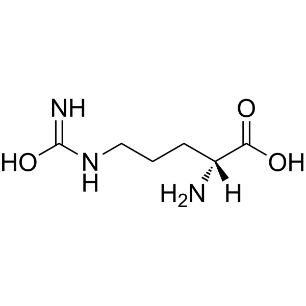
血管紧张素Ⅱ
Angiotensin II (Angiotensin II) 是一种血管收缩剂,是肾素/血管紧张素系统的主要生物活性肽。Angiotensin II human 在调节人类血压中起着核心作用,主要通过血管紧张素 II 与 G 蛋白偶联受体 (GPCRs)、血管紧张素II 1型受体 (AT1R) 和血管紧张素II 2型受体 (AT2R) 之间的相互作用来介导。Angiotensin II human 刺激交感神经刺激,增加醛固酮生物合成和肾脏活动。Angiotensin II human 诱导血管平滑肌细胞生长,增加成纤维细胞中 I 型和 III 型胶原的合成,导致血管壁和心肌增厚,并导致纤维化。Angiotensin II human 也诱导细胞凋亡 (apoptosis)。Angiotensin II human 通过 LOX-1 依赖的氧化还原敏感途径诱导内皮细胞毛细血管形成。
Western blot analysis of extracts from THP-1(lane 2(20μg), Jurkat (lane 3(20μg) and NIH3T3(lane 4(20μg) using FOXO1A (HY-P80132) Rabbit mAb. Proteins were transferred
to a PVDF membrane and blocked with 5% non-fat milk in TBST for 2 hour at room temperature. The primary antibody (1/1000) and Loading control antibody (Beta Actin, HY-P80438, 1/10000) was
used in 5% non-fat milk in TBST at 4°C overnight. Goat Anti-Mouse/Rabbit IgG-HRP Secondary Antibody (1/10000) was used for 1 hour at room temperature.
Western blot analysis of extracts from THP-1(lane 2(20μg), Jurkat (lane 3(20μg) and NIH3T3(lane 4(20μg) using FOXO1A (HY-P80132) Rabbit mAb. Proteins were transferred
to a PVDF membrane and blocked with 5% non-fat milk in TBST for 2 hour at room temperature. The primary antibody (1/1000) and Loading control antibody (Beta Actin, HY-P80438, 1/10000) was
MedchemExpress Validation 03
Western blot analysis of extracts from THP-1(lane 2(20μg), Jurkat (lane 3(20μg) and NIH3T3(lane 4(20μg) using FOXO1A (HY-P80132) Rabbit mAb. Proteins were transferred
MedchemExpress Validation 04
Western blot analysis of extracts from THP-1(lane 2(20μg), Jurkat (lane 3(20μg) and NIH3T3(lane 4(20μg) using FOXO1A (HY-P80132) Rabbit mAb. Proteins were transferred
to a PVDF membrane and blocked with 5% non-fat milk in TBST for 2 hour at room temperature. The primary antibody (1/1000) and Loading control antibody (Beta Actin, HY-P80438, 1/10000) was
used in 5% non-fat milk in TBST at 4°C overnight. Goat Anti-Mouse/Rabbit IgG-HRP Secondary Antibody (1/10000) was used for 1 hour at room temperature.
MedchemExpress Validation
Western blot analysis of extracts from THP-1(lane 2(20μg), Jurkat (lane 3(20μg) and NIH3T3(lane 4(20μg) using FOXO1A (HY-P80132) Rabbit mAb. Proteins were transferred
to a PVDF membrane and blocked with 5% non-fat milk in TBST for 2 hour at room temperature. The primary antibody (1/1000) and Loading control antibody (Beta Actin, HY-P80438, 1/10000) was
used in 5% non-fat milk in TBST at 4°C overnight. Goat Anti-Mouse/Rabbit IgG-HRP Secondary Antibody (1/10000) was used for 1 hour at room temperature.
MedchemExpress Validation
Western blot analysis of extracts from THP-1(lane 2(20μg), Jurkat (lane 3(20μg) and NIH3T3(lane 4(20μg) using FOXO1A (HY-P80132) Rabbit mAb. Proteins were transferred
to a PVDF membrane and blocked with 5% non-fat milk in TBST for 2 hour at room temperature. The primary antibody (1/1000) and Loading control antibody (Beta Actin, HY-P80438, 1/10000) was
used in 5% non-fat milk in TBST at 4°C overnight. Goat Anti-Mouse/Rabbit IgG-HRP Secondary Antibody (1/10000) was used for 1 hour at room temperature.