Myeloperoxidase, Human Neutrophil 是一种过氧化物酶。Myeloperoxidase, Human Neutrophil 是一种强效抗菌剂,通过催化依赖 H2O2 的氯离子氧化来生成次氯酸。Myeloperoxidase, Human Neutrophil 催化 N-视黄基-亚胺-N-视黄基乙醇胺 (一种有毒形式的视网膜脂褐素) 的降解。Myeloperoxidase, Human Neutrophil 还会引发溶酶体应激和细胞死亡。Myeloperoxidase, Human Neutrophil 可用于炎症和感染研究。
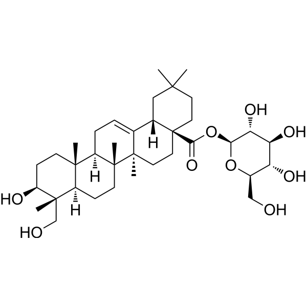
Ganoleucoin R 是一种可以从 Ganoderma leucocontextum 中分离得到的三萜类化合物。Ganoleucoin R 对 H2O2 诱导的氧化损伤具有保护作用,也能促进 PC12 细胞神经突生长。Ganoleucoin R 具有神经保护和促神经发生的活性,可用于神经退行性疾病的研究。
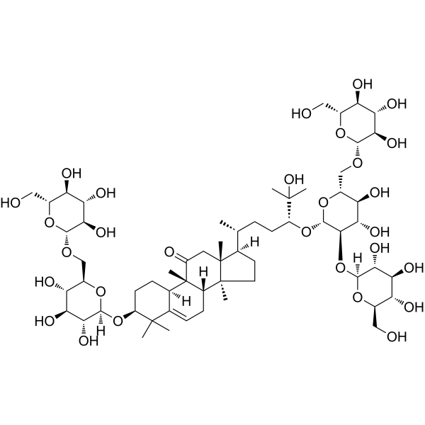
11-氧-罗汉果苷 V (标准品)
11-oxo-mogroside V (Standard) 是 11-oxo-mogroside V 的分析标准品。本产品用于研究及分析应用。11-oxo-mogroside V 是一种天然甜味剂,具有很强的抗氧化活性。11-oxo-mogroside V 对活性氧物质具有显着的抑制作用,作用于 O2-,H2O2 和 *OH,EC50 分别为 4.79,16.52 和 146.17 μg/mL。
吉奥诺苷D
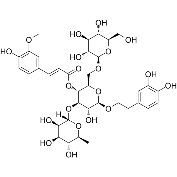
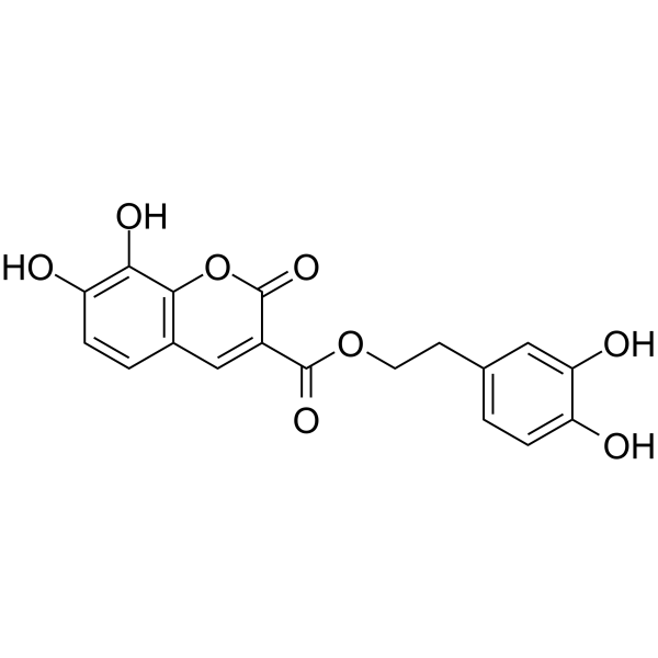
Jionoside D 是一种具有抗氧化作用的羟基肉桂酸酯。Jionoside D 具有清除胞内活性氧 (ROS) 和 DPPH 自由基的活性,以及抑制脂质过氧化的活性。Jionoside D 可降低 H2O2 诱导的 V79-4 细胞凋亡 (apoptosis)。Jionoside D 可提高细胞抗氧化酶、SOD 和过氧化氢酶的活性。
Oxalate oxidase, B. subtilis (EC 1.2.3.4) 是一种作用于供体醛基或羰基,以氧为受体的氧化还原酶。Oxalate oxidase, B 参与乙醛酸和二羧酸的代谢。Oxalate oxidase, B 有两种辅因子:FAD 和锰。Oxalate oxidase, B 的三种底物是草酸、O2 和 H+,而其两种产物是 CO2 和 H2O2。
麦冬皂苷D
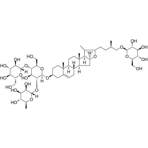
Ophiopogonin D 是可以从麦冬 (Ophiopogon japonicus) 的块茎中分离的,是一种罕见的天然存在的 C29 甾体糖苷。Ophiopogonin D 是 CYP2J3 诱导剂,其通过增加人脐静脉内皮细胞 (HUVECs) 中 CYP2J2/EETs 和 PPARα 的表达,显着抑制 Ang II 诱导的 NF-κB 核转位,IκBα 下调,细胞内 Ca2+ 过载和促炎细胞因子的激活。Ophiopogonin D 能抑制 RAW264.7 细胞的破骨细胞分化。Ophiopogonin D 作为抗氧化剂,在过氧化氢 (H2O2) 诱导的内皮损伤中具有保护作用。Ophiopogonin D 能阻断 ERK 信号级联。Ophiopogonin D 可缓解高脂饮食引起的代谢综合征,并改变小鼠肠道菌群的结构。Ophiopogonin D 已被用于炎症、代谢和心血管疾病。
麦冬皂苷D (标准品)
Ophiopogonin D (Standard) 是 Ophiopogonin D 的分析标准品。本产品用于研究及分析应用。Ophiopogonin D 是可以从麦冬 (Ophiopogon japonicus) 的块茎中分离的,是一种罕见的天然存在的 C29 甾体糖苷。Ophiopogonin D 是 CYP2J3 诱导剂,其通过增加人脐静脉内皮细胞 (HUVECs) 中 CYP2J2/EETs 和 PPARα 的表达,显着抑制 Ang II 诱导的 NF-κB 核转位,IκBα 下调,细胞内 Ca2+ 过载和促炎细胞因子的激活。Ophiopogonin D 能抑制 RAW264.7 细胞的破骨细胞分化。Ophiopogonin D 作为抗氧化剂,在过氧化氢 (H2O2) 诱导的内皮损伤中具有保护作用。Ophiopogonin D 能阻断 ERK 信号级联。Ophiopogonin D 可缓解高脂饮食引起的代谢综合征,并改变小鼠肠道菌群的结构。Ophiopogonin D 已被用于炎症、代谢和心血管疾病。
麦冬皂苷D
Ophiopogonin D 是可以从麦冬 (Ophiopogon japonicus) 的块茎中分离的,是一种罕见的天然存在的 C29 甾体糖苷。Ophiopogonin D 是 CYP2J3 诱导剂,其通过增加人脐静脉内皮细胞 (HUVECs) 中 CYP2J2/EETs 和 PPARα 的表达,显着抑制 Ang II 诱导的 NF-κB 核转位,IκBα 下调,细胞内 Ca2+ 过载和促炎细胞因子的激活。Ophiopogonin D 能抑制 RAW264.7 细胞的破骨细胞分化。Ophiopogonin D 作为抗氧化剂,在过氧化氢 (H2O2) 诱导的内皮损伤中具有保护作用。Ophiopogonin D 能阻断 ERK 信号级联。Ophiopogonin D 可缓解高脂饮食引起的代谢综合征,并改变小鼠肠道菌群的结构。Ophiopogonin D 已被用于炎症、代谢和心血管疾病。
麦冬皂苷D (标准品)
Ophiopogonin D (Standard) 是 Ophiopogonin D 的分析标准品。本产品用于研究及分析应用。Ophiopogonin D 是可以从麦冬 (Ophiopogon japonicus) 的块茎中分离的,是一种罕见的天然存在的 C29 甾体糖苷。Ophiopogonin D 是 CYP2J3 诱导剂,其通过增加人脐静脉内皮细胞 (HUVECs) 中 CYP2J2/EETs 和 PPARα 的表达,显着抑制 Ang II 诱导的 NF-κB 核转位,IκBα 下调,细胞内 Ca2+ 过载和促炎细胞因子的激活。Ophiopogonin D 能抑制 RAW264.7 细胞的破骨细胞分化。Ophiopogonin D 作为抗氧化剂,在过氧化氢 (H2O2) 诱导的内皮损伤中具有保护作用。Ophiopogonin D 能阻断 ERK 信号级联。Ophiopogonin D 可缓解高脂饮食引起的代谢综合征,并改变小鼠肠道菌群的结构。Ophiopogonin D 已被用于炎症、代谢和心血管疾病。
Ganoleucoin R 是一种可以从 Ganoderma leucocontextum 中分离得到的三萜类化合物。Ganoleucoin R 对 H2O2 诱导的氧化损伤具有保护作用,也能促进 PC12 细胞神经突生长。Ganoleucoin R 具有神经保护和促神经发生的活性,可用于神经退行性疾病的研究。
11-氧-罗汉果苷 V (标准品)
11-oxo-mogroside V (Standard) 是 11-oxo-mogroside V 的分析标准品。本产品用于研究及分析应用。11-oxo-mogroside V 是一种天然甜味剂,具有很强的抗氧化活性。11-oxo-mogroside V 对活性氧物质具有显着的抑制作用,作用于 O2-,H2O2 和 *OH,EC50 分别为 4.79,16.52 和 146.17 μg/mL。
吉奥诺苷D
Jionoside D 是一种具有抗氧化作用的羟基肉桂酸酯。Jionoside D 具有清除胞内活性氧 (ROS) 和 DPPH 自由基的活性,以及抑制脂质过氧化的活性。Jionoside D 可降低 H2O2 诱导的 V79-4 细胞凋亡 (apoptosis)。Jionoside D 可提高细胞抗氧化酶、SOD 和过氧化氢酶的活性。
Western blot analysis of extracts from THP-1(lane 2(20μg), Jurkat (lane 3(20μg) and NIH3T3(lane 4(20μg) using FOXO1A (HY-P80132) Rabbit mAb. Proteins were transferred
to a PVDF membrane and blocked with 5% non-fat milk in TBST for 2 hour at room temperature. The primary antibody (1/1000) and Loading control antibody (Beta Actin, HY-P80438, 1/10000) was
used in 5% non-fat milk in TBST at 4°C overnight. Goat Anti-Mouse/Rabbit IgG-HRP Secondary Antibody (1/10000) was used for 1 hour at room temperature.
Western blot analysis of extracts from THP-1(lane 2(20μg), Jurkat (lane 3(20μg) and NIH3T3(lane 4(20μg) using FOXO1A (HY-P80132) Rabbit mAb. Proteins were transferred
to a PVDF membrane and blocked with 5% non-fat milk in TBST for 2 hour at room temperature. The primary antibody (1/1000) and Loading control antibody (Beta Actin, HY-P80438, 1/10000) was
MedchemExpress Validation 03
Western blot analysis of extracts from THP-1(lane 2(20μg), Jurkat (lane 3(20μg) and NIH3T3(lane 4(20μg) using FOXO1A (HY-P80132) Rabbit mAb. Proteins were transferred
MedchemExpress Validation 04
Western blot analysis of extracts from THP-1(lane 2(20μg), Jurkat (lane 3(20μg) and NIH3T3(lane 4(20μg) using FOXO1A (HY-P80132) Rabbit mAb. Proteins were transferred
to a PVDF membrane and blocked with 5% non-fat milk in TBST for 2 hour at room temperature. The primary antibody (1/1000) and Loading control antibody (Beta Actin, HY-P80438, 1/10000) was
used in 5% non-fat milk in TBST at 4°C overnight. Goat Anti-Mouse/Rabbit IgG-HRP Secondary Antibody (1/10000) was used for 1 hour at room temperature.
MedchemExpress Validation
Western blot analysis of extracts from THP-1(lane 2(20μg), Jurkat (lane 3(20μg) and NIH3T3(lane 4(20μg) using FOXO1A (HY-P80132) Rabbit mAb. Proteins were transferred
to a PVDF membrane and blocked with 5% non-fat milk in TBST for 2 hour at room temperature. The primary antibody (1/1000) and Loading control antibody (Beta Actin, HY-P80438, 1/10000) was
used in 5% non-fat milk in TBST at 4°C overnight. Goat Anti-Mouse/Rabbit IgG-HRP Secondary Antibody (1/10000) was used for 1 hour at room temperature.
MedchemExpress Validation
Western blot analysis of extracts from THP-1(lane 2(20μg), Jurkat (lane 3(20μg) and NIH3T3(lane 4(20μg) using FOXO1A (HY-P80132) Rabbit mAb. Proteins were transferred
to a PVDF membrane and blocked with 5% non-fat milk in TBST for 2 hour at room temperature. The primary antibody (1/1000) and Loading control antibody (Beta Actin, HY-P80438, 1/10000) was
used in 5% non-fat milk in TBST at 4°C overnight. Goat Anti-Mouse/Rabbit IgG-HRP Secondary Antibody (1/10000) was used for 1 hour at room temperature.