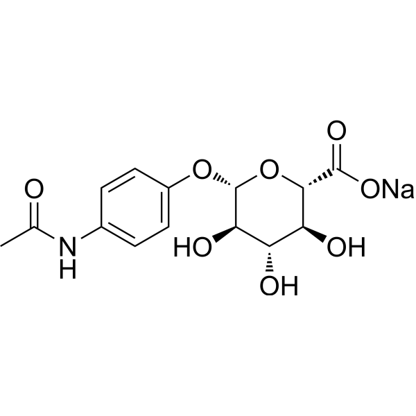
吲哚菁绿 (标准品)
Indocyanine green (Standard)是 Indocyanine green 的分析标准品。本产品用于研究及分析应用。Indocyanine green (Foxgreen) 是一种低毒性荧光剂,广泛用于医学诊断,如确定心输出量,肝功能和肝血流量,以及用于眼科血管造影。
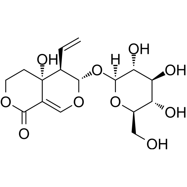
Cuneataside E 是一种苯丙素苷类化合物,发现于截叶铁扫帚 (Lespedeza cuneata)。
Cuneataside E 对 N-acetyl-p-aminophenol (APAP) 诱导的人肝癌细胞系 HepG2 的毒性有保护作用。Cuneataside E 有望用于肝脏疾病的研究。
脂质X
Lipid X 是一种作为脂质 A 单糖前体的 2,3-二酰基葡萄糖胺 -1-磷酸,兼具 LPS 拮抗剂与弱激动剂活性。Lipid X 能通过抑制肿瘤坏死因子产生、单核细胞促凝活性及中性粒细胞启动来发挥保护作用。Lipid X 在实验动物中可能诱发短暂的肺动脉高压、中性粒细胞减少及轻度致热效应。Lipid X 的毒性较低且无体外抗菌活性,但显著降低革兰氏阴性菌感染和内毒素暴露后的死亡率。Lipid X 易在肝组织蓄积并与循环细胞成分结合,还可通过转酯作用转化为脂质 Y。Lipid X 可用于革兰氏阴性菌败血症、内毒素血症及相关肺动脉高压的研究。
DILI 已被证实是一个由多种机制参与的复杂病理过程,其中包括肝脏的结构和功能完整性的直接损害(例如线粒体功能障碍);产生改变肝细胞结构和功能的代谢产物;产生能与肝蛋白结合的反应性药物代谢产物,从而产生新的抗原性药物-蛋白质加合物,这些新的加合物被宿主的防御系统所针对(半抗原假说),并引发损害肝脏的全身超敏反应(即药物过敏)。
MCE 肝脏毒性化合物库包含 640 种肝脏毒性化合物,是进行肝损伤及相关药物毒理研究的有力工具,该化合物库也可以用来探究 DILI 发病机制,识别早期 DILI 标记物,在药物开发中及时发现并规避可能诱导肝损伤的药物设计,有助于药物的顺利上市。
吲哚菁绿 (标准品)
Indocyanine green (Standard)是 Indocyanine green 的分析标准品。本产品用于研究及分析应用。Indocyanine green (Foxgreen) 是一种低毒性荧光剂,广泛用于医学诊断,如确定心输出量,肝功能和肝血流量,以及用于眼科血管造影。
Cuneataside E 是一种苯丙素苷类化合物,发现于截叶铁扫帚 (Lespedeza cuneata)。
Cuneataside E 对 N-acetyl-p-aminophenol (APAP) 诱导的人肝癌细胞系 HepG2 的毒性有保护作用。Cuneataside E 有望用于肝脏疾病的研究。
Western blot analysis of extracts from THP-1(lane 2(20μg), Jurkat (lane 3(20μg) and NIH3T3(lane 4(20μg) using FOXO1A (HY-P80132) Rabbit mAb. Proteins were transferred
to a PVDF membrane and blocked with 5% non-fat milk in TBST for 2 hour at room temperature. The primary antibody (1/1000) and Loading control antibody (Beta Actin, HY-P80438, 1/10000) was
used in 5% non-fat milk in TBST at 4°C overnight. Goat Anti-Mouse/Rabbit IgG-HRP Secondary Antibody (1/10000) was used for 1 hour at room temperature.
Western blot analysis of extracts from THP-1(lane 2(20μg), Jurkat (lane 3(20μg) and NIH3T3(lane 4(20μg) using FOXO1A (HY-P80132) Rabbit mAb. Proteins were transferred
to a PVDF membrane and blocked with 5% non-fat milk in TBST for 2 hour at room temperature. The primary antibody (1/1000) and Loading control antibody (Beta Actin, HY-P80438, 1/10000) was
MedchemExpress Validation 03
Western blot analysis of extracts from THP-1(lane 2(20μg), Jurkat (lane 3(20μg) and NIH3T3(lane 4(20μg) using FOXO1A (HY-P80132) Rabbit mAb. Proteins were transferred
MedchemExpress Validation 04
Western blot analysis of extracts from THP-1(lane 2(20μg), Jurkat (lane 3(20μg) and NIH3T3(lane 4(20μg) using FOXO1A (HY-P80132) Rabbit mAb. Proteins were transferred
to a PVDF membrane and blocked with 5% non-fat milk in TBST for 2 hour at room temperature. The primary antibody (1/1000) and Loading control antibody (Beta Actin, HY-P80438, 1/10000) was
used in 5% non-fat milk in TBST at 4°C overnight. Goat Anti-Mouse/Rabbit IgG-HRP Secondary Antibody (1/10000) was used for 1 hour at room temperature.
MedchemExpress Validation
Western blot analysis of extracts from THP-1(lane 2(20μg), Jurkat (lane 3(20μg) and NIH3T3(lane 4(20μg) using FOXO1A (HY-P80132) Rabbit mAb. Proteins were transferred
to a PVDF membrane and blocked with 5% non-fat milk in TBST for 2 hour at room temperature. The primary antibody (1/1000) and Loading control antibody (Beta Actin, HY-P80438, 1/10000) was
used in 5% non-fat milk in TBST at 4°C overnight. Goat Anti-Mouse/Rabbit IgG-HRP Secondary Antibody (1/10000) was used for 1 hour at room temperature.
MedchemExpress Validation
Western blot analysis of extracts from THP-1(lane 2(20μg), Jurkat (lane 3(20μg) and NIH3T3(lane 4(20μg) using FOXO1A (HY-P80132) Rabbit mAb. Proteins were transferred
to a PVDF membrane and blocked with 5% non-fat milk in TBST for 2 hour at room temperature. The primary antibody (1/1000) and Loading control antibody (Beta Actin, HY-P80438, 1/10000) was
used in 5% non-fat milk in TBST at 4°C overnight. Goat Anti-Mouse/Rabbit IgG-HRP Secondary Antibody (1/10000) was used for 1 hour at room temperature.