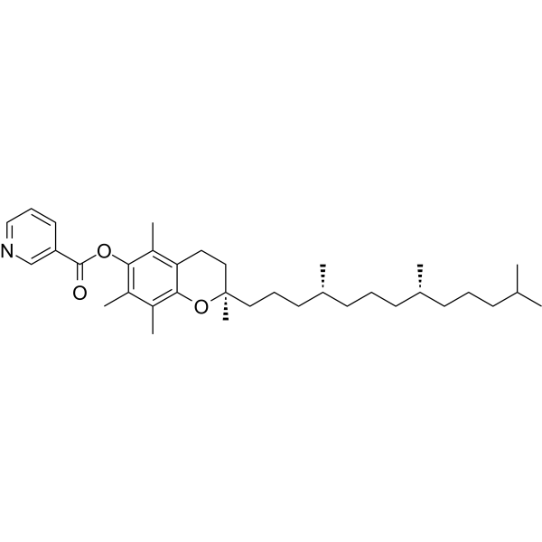
Roridin E 是一种葡萄糖-6-磷酸酶 (G6Pase) 抑制剂和抗生素,是 Roridin A (HY-N9599) 的代谢副产物。Roridin E 会引发显著的氧化应激反应,表现为消耗体内谷胱甘肽、诱导肝脏脂质过氧化以及抑制肾脏超氧化物歧化酶活性。Roridin E 能够降低大鼠血糖水平,但具有急性毒性 (与亚油酸 (HY-N0729) 联用时毒性增强),并会导致雄性白化小鼠出现肝毒性。Roridin E 可诱导血液总蛋白降低以及总脂质、γ-谷氨酰转移酶、碱性磷酸酶和 5'-核苷酸酶水平升高。Roridin E 能够从霉菌中分离得到,具有与 Verrucarin A (HY-107426) 和 Roridin A 相似的细胞抑制和真菌抑制活性。Roridin E 在啮齿类动物中具有体内活性,常被用于肝毒性相关研究。
莱苞迪甙 A (标准品)
Rebaudioside A (Standard) 是 Rebaudioside A 的分析标准品。本产品用于研究及分析应用。Rebaudioside A 是一种口服有效的甜菊醇糖苷,具有较高的甜味。Rebaudioside A 是 α-glucosidase 抑制剂,IC50 值是 35.01 μg/mL。Rebaudioside A 通过葡萄糖依赖性的方式,增加 β 细胞内的 ATP/ADP 比值,从而抑制 KATP 通道,导致细胞膜去极化、钙离子内流,最终刺激胰岛素分泌。Rebaudioside A 通过抑制胆固醇合成的限速酶 HMGCR,激活 SREBP 信号通路,导致细胞表面 LDLR 表达增加,从而促进血液中 LDL-C 的摄取。Rebaudioside A 可用于研究血糖、血脂调节和抗肥胖。
番茄皂苷A
Esculeoside A 是一种具有口服活性的螺甾烷型甾体生物碱糖苷,存在于 Lycopersicon esculentum var. cerasiforme 和 Lycopersicon esculentum 的成熟果实中。Esculeoside A 是一种心脏保护剂和降糖剂。Esculeoside A 可调控心脏组织中的 NF-κB、Nrf2/Keap1 信号通路,减轻炎症反应与凋亡 (apoptosis),阻止心肌细胞肥大,并降低血清脂质水平。Esculeoside A 可通过调控 IRS-1、GCK、AMPK 和 PEPCK 的表达来调节糖代谢,降低空腹血糖,改善葡萄糖耐量。Esculeoside A 可抑制 TLR4/p-NFκB 信号通路,从而阻碍树突状细胞成熟及异体 T 细胞增殖,并抑制乳腺癌与黑色素瘤细胞的生长。Esculeoside A 可抑制 ACAT 活性,减少巨噬细胞中胆固醇酯的积累。Esculeoside A 可用于糖尿病心肌病、2 型糖尿病、特应性皮炎、肿瘤及动脉粥样硬化性疾病的研究。
MCE 油红 O 染色试剂盒 (细胞涂片专用) 可有效染色各种大小的脂滴,包括较小的脂滴,且能够从溶剂中优先吸附染料。适用于细胞涂片、骨髓涂片、体液涂片、血液涂片等的油红 O 染色。使用时,标本不应采用含有乙醇的固定液。若需要固定,可使用 10% 福尔马林。脂肪的阳性染色结果通常呈橘黄色至红色,但具体颜色会根据脂质浓度有所不同。
Roridin E 是一种葡萄糖-6-磷酸酶 (G6Pase) 抑制剂和抗生素,是 Roridin A (HY-N9599) 的代谢副产物。Roridin E 会引发显著的氧化应激反应,表现为消耗体内谷胱甘肽、诱导肝脏脂质过氧化以及抑制肾脏超氧化物歧化酶活性。Roridin E 能够降低大鼠血糖水平,但具有急性毒性 (与亚油酸 (HY-N0729) 联用时毒性增强),并会导致雄性白化小鼠出现肝毒性。Roridin E 可诱导血液总蛋白降低以及总脂质、γ-谷氨酰转移酶、碱性磷酸酶和 5'-核苷酸酶水平升高。Roridin E 能够从霉菌中分离得到,具有与 Verrucarin A (HY-107426) 和 Roridin A 相似的细胞抑制和真菌抑制活性。Roridin E 在啮齿类动物中具有体内活性,常被用于肝毒性相关研究。
莱苞迪甙 A (标准品)
Rebaudioside A (Standard) 是 Rebaudioside A 的分析标准品。本产品用于研究及分析应用。Rebaudioside A 是一种口服有效的甜菊醇糖苷,具有较高的甜味。Rebaudioside A 是 α-glucosidase 抑制剂,IC50 值是 35.01 μg/mL。Rebaudioside A 通过葡萄糖依赖性的方式,增加 β 细胞内的 ATP/ADP 比值,从而抑制 KATP 通道,导致细胞膜去极化、钙离子内流,最终刺激胰岛素分泌。Rebaudioside A 通过抑制胆固醇合成的限速酶 HMGCR,激活 SREBP 信号通路,导致细胞表面 LDLR 表达增加,从而促进血液中 LDL-C 的摄取。Rebaudioside A 可用于研究血糖、血脂调节和抗肥胖。
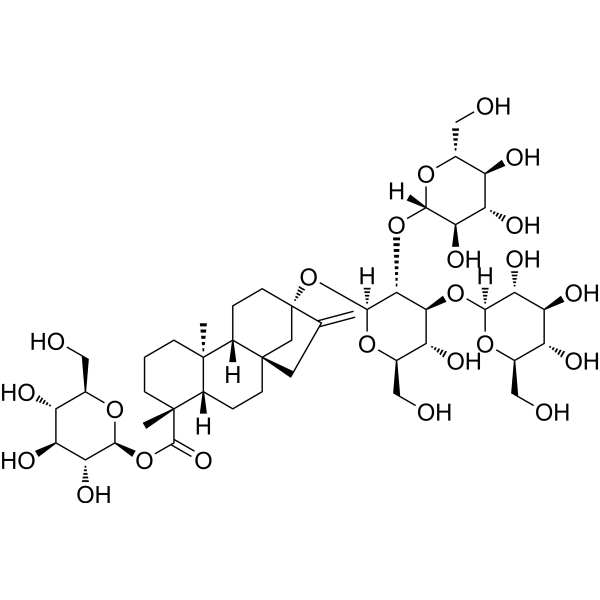
番茄皂苷A
Esculeoside A 是一种具有口服活性的螺甾烷型甾体生物碱糖苷,存在于 Lycopersicon esculentum var. cerasiforme 和 Lycopersicon esculentum 的成熟果实中。Esculeoside A 是一种心脏保护剂和降糖剂。Esculeoside A 可调控心脏组织中的 NF-κB、Nrf2/Keap1 信号通路,减轻炎症反应与凋亡 (apoptosis),阻止心肌细胞肥大,并降低血清脂质水平。Esculeoside A 可通过调控 IRS-1、GCK、AMPK 和 PEPCK 的表达来调节糖代谢,降低空腹血糖,改善葡萄糖耐量。Esculeoside A 可抑制 TLR4/p-NFκB 信号通路,从而阻碍树突状细胞成熟及异体 T 细胞增殖,并抑制乳腺癌与黑色素瘤细胞的生长。Esculeoside A 可抑制 ACAT 活性,减少巨噬细胞中胆固醇酯的积累。Esculeoside A 可用于糖尿病心肌病、2 型糖尿病、特应性皮炎、肿瘤及动脉粥样硬化性疾病的研究。
Western blot analysis of extracts from THP-1(lane 2(20μg), Jurkat (lane 3(20μg) and NIH3T3(lane 4(20μg) using FOXO1A (HY-P80132) Rabbit mAb. Proteins were transferred
to a PVDF membrane and blocked with 5% non-fat milk in TBST for 2 hour at room temperature. The primary antibody (1/1000) and Loading control antibody (Beta Actin, HY-P80438, 1/10000) was
used in 5% non-fat milk in TBST at 4°C overnight. Goat Anti-Mouse/Rabbit IgG-HRP Secondary Antibody (1/10000) was used for 1 hour at room temperature.
Western blot analysis of extracts from THP-1(lane 2(20μg), Jurkat (lane 3(20μg) and NIH3T3(lane 4(20μg) using FOXO1A (HY-P80132) Rabbit mAb. Proteins were transferred
to a PVDF membrane and blocked with 5% non-fat milk in TBST for 2 hour at room temperature. The primary antibody (1/1000) and Loading control antibody (Beta Actin, HY-P80438, 1/10000) was
MedchemExpress Validation 03
Western blot analysis of extracts from THP-1(lane 2(20μg), Jurkat (lane 3(20μg) and NIH3T3(lane 4(20μg) using FOXO1A (HY-P80132) Rabbit mAb. Proteins were transferred
MedchemExpress Validation 04
Western blot analysis of extracts from THP-1(lane 2(20μg), Jurkat (lane 3(20μg) and NIH3T3(lane 4(20μg) using FOXO1A (HY-P80132) Rabbit mAb. Proteins were transferred
to a PVDF membrane and blocked with 5% non-fat milk in TBST for 2 hour at room temperature. The primary antibody (1/1000) and Loading control antibody (Beta Actin, HY-P80438, 1/10000) was
used in 5% non-fat milk in TBST at 4°C overnight. Goat Anti-Mouse/Rabbit IgG-HRP Secondary Antibody (1/10000) was used for 1 hour at room temperature.
MedchemExpress Validation
Western blot analysis of extracts from THP-1(lane 2(20μg), Jurkat (lane 3(20μg) and NIH3T3(lane 4(20μg) using FOXO1A (HY-P80132) Rabbit mAb. Proteins were transferred
to a PVDF membrane and blocked with 5% non-fat milk in TBST for 2 hour at room temperature. The primary antibody (1/1000) and Loading control antibody (Beta Actin, HY-P80438, 1/10000) was
used in 5% non-fat milk in TBST at 4°C overnight. Goat Anti-Mouse/Rabbit IgG-HRP Secondary Antibody (1/10000) was used for 1 hour at room temperature.
MedchemExpress Validation
Western blot analysis of extracts from THP-1(lane 2(20μg), Jurkat (lane 3(20μg) and NIH3T3(lane 4(20μg) using FOXO1A (HY-P80132) Rabbit mAb. Proteins were transferred
to a PVDF membrane and blocked with 5% non-fat milk in TBST for 2 hour at room temperature. The primary antibody (1/1000) and Loading control antibody (Beta Actin, HY-P80438, 1/10000) was
used in 5% non-fat milk in TBST at 4°C overnight. Goat Anti-Mouse/Rabbit IgG-HRP Secondary Antibody (1/10000) was used for 1 hour at room temperature.