Defensin HNP-1 human 是一种人中性粒细胞肽 (HNPs)。Defensin HNP-1 human 具有免疫调节功能,可延迟中性粒细胞凋亡 (apoptosis)。Defensin HNP-1 human 通过抑制 DNA/RNA/蛋白质合成,干扰代谢途径,具有广泛的抗菌活性。Defensin HNP-1 human 对 HIV、HSV-1、HSV-2、CMV、流感病毒等有直接灭活作用。Defensin HNP-1 human 具有抗利什曼原虫活性。Defensin HNP-1 human 参与早期动脉粥样硬化发展时的内皮细胞功能障碍。
髓过氧化物酶
Myeloperoxidase, human white blood cells (MPO) 是一种过氧化物酶。Myeloperoxidase, human white blood cells 通过促进活性氧 (ROS) 和活性氮 (RNS) 的产生,调节小胶质细胞和中性粒细胞的极化和炎症相关信号通路来介导氧化应激。Myeloperoxidase, human white blood cells 具有抗菌 (antibacterial) 活性。
Defensin HNP-1 human TFA 是一种人中性粒细胞肽 (HNPs)。Defensin HNP-1 human TFA 具有免疫调节功能,可延迟中性粒细胞凋亡 (apoptosis)。Defensin HNP-1 human TFA 通过抑制 DNA/RNA/蛋白质合成,干扰代谢途径,具有广泛的抗菌活性。Defensin HNP-1 human TFA 对 HIV、HSV-1、HSV-2、CMV、流感病毒等有直接灭活作用。Defensin HNP-1 human TFA 具有抗利什曼原虫活性。Defensin HNP-1 human TFA 参与早期动脉粥样硬化发展时的内皮细胞功能障碍。
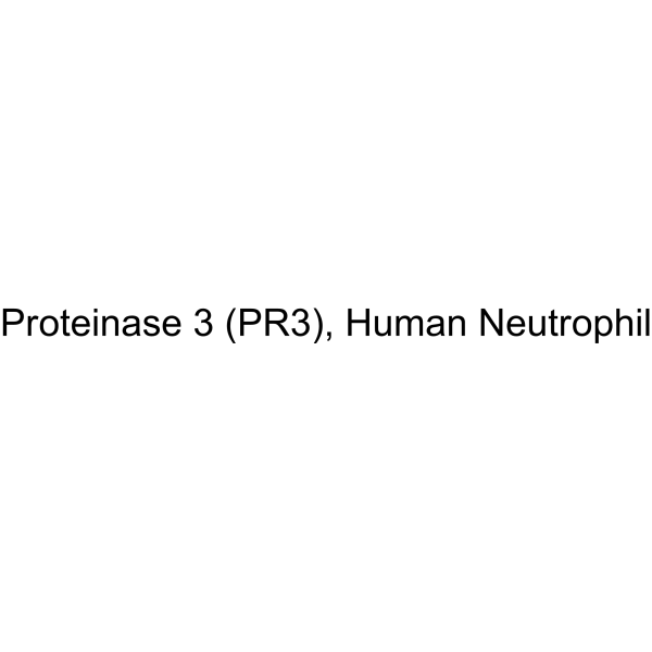
弹性蛋白酶来源于人类白血球
Elastase, Human leukocytes 是一种丝氨酸蛋白酶,存在于中性粒细胞的亲氮颗粒中。Elastase, Human leukocytes 潜在底物包括细胞外基质的几乎所有成分,以及凝血因子、补体、免疫球蛋白和细胞因子等多种蛋白质,有强大的蛋白质水解功能,参与炎症组织损伤的发病机制。
Myeloperoxidase, Human Neutrophil 是一种过氧化物酶。Myeloperoxidase, Human Neutrophil 是一种强效抗菌剂,通过催化依赖 H2O2 的氯离子氧化来生成次氯酸。Myeloperoxidase, Human Neutrophil 催化 N-视黄基-亚胺-N-视黄基乙醇胺 (一种有毒形式的视网膜脂褐素) 的降解。Myeloperoxidase, Human Neutrophil 还会引发溶酶体应激和细胞死亡。Myeloperoxidase, Human Neutrophil 可用于炎症和感染研究。
知母皂苷IA
Anemarrhenasaponin Ia 是一种甾体皂苷。Anemarrhenasaponin Ia 可从知母 (Anemarrhena asphodeloides Bunge) 根茎中分离得到。Anemarrhenasaponin Ia 可抑制血小板聚集。Anemarrhenasaponin Ia 诱导轻微的浓度依赖性溶血。Anemarrhenasaponin Ia 可抑制 fMLP 和 AA 诱导的人中性粒细胞超氧阴离子生成,同时增强 PMA 诱导的人中性粒细胞超氧阴离子生成。Anemarrhenasaponin Ia 可用于血栓形成的相关研究。。
Human CCL22 mRNA 编码人类 CC 基序趋化因子配体 22 (CCL22) 蛋白,该蛋白是一种对单核细胞、树突状细胞、自然杀伤细胞和慢性活化 T 淋巴细胞具有趋化活性的细胞因子。CCL22 对原代活化 T 淋巴细胞也表现出轻微的趋化活性,但对中性粒细胞、嗜酸性粒细胞和静息 T 淋巴细胞没有趋化活性。
APO-15 是一种磷脂酰丝氨酸结合型荧光探针及凋亡成像试剂。APO-15 在蛋白水解和氧化条件下具有较高的化学稳定性,可在临床前小鼠模型中实现药物诱导凋亡的定量与成像,且适用于固定组织样本及多种 in vivo 给药途径(Ex = 488 nm; Em = 525 nm) 。APO-15 可用于急性肺损伤及乳腺癌的相关研究。
Defensin HNP-1 human 是一种人中性粒细胞肽 (HNPs)。Defensin HNP-1 human 具有免疫调节功能,可延迟中性粒细胞凋亡 (apoptosis)。Defensin HNP-1 human 通过抑制 DNA/RNA/蛋白质合成,干扰代谢途径,具有广泛的抗菌活性。Defensin HNP-1 human 对 HIV、HSV-1、HSV-2、CMV、流感病毒等有直接灭活作用。Defensin HNP-1 human 具有抗利什曼原虫活性。Defensin HNP-1 human 参与早期动脉粥样硬化发展时的内皮细胞功能障碍。
Defensin HNP-1 human TFA 是一种人中性粒细胞肽 (HNPs)。Defensin HNP-1 human TFA 具有免疫调节功能,可延迟中性粒细胞凋亡 (apoptosis)。Defensin HNP-1 human TFA 通过抑制 DNA/RNA/蛋白质合成,干扰代谢途径,具有广泛的抗菌活性。Defensin HNP-1 human TFA 对 HIV、HSV-1、HSV-2、CMV、流感病毒等有直接灭活作用。Defensin HNP-1 human TFA 具有抗利什曼原虫活性。Defensin HNP-1 human TFA 参与早期动脉粥样硬化发展时的内皮细胞功能障碍。
APO-15 是一种磷脂酰丝氨酸结合型荧光探针及凋亡成像试剂。APO-15 在蛋白水解和氧化条件下具有较高的化学稳定性,可在临床前小鼠模型中实现药物诱导凋亡的定量与成像,且适用于固定组织样本及多种 in vivo 给药途径(Ex = 488 nm; Em = 525 nm) 。APO-15 可用于急性肺损伤及乳腺癌的相关研究。
知母皂苷IA
Anemarrhenasaponin Ia 是一种甾体皂苷。Anemarrhenasaponin Ia 可从知母 (Anemarrhena asphodeloides Bunge) 根茎中分离得到。Anemarrhenasaponin Ia 可抑制血小板聚集。Anemarrhenasaponin Ia 诱导轻微的浓度依赖性溶血。Anemarrhenasaponin Ia 可抑制 fMLP 和 AA 诱导的人中性粒细胞超氧阴离子生成,同时增强 PMA 诱导的人中性粒细胞超氧阴离子生成。Anemarrhenasaponin Ia 可用于血栓形成的相关研究。。
Human CCL22 mRNA 编码人类 CC 基序趋化因子配体 22 (CCL22) 蛋白,该蛋白是一种对单核细胞、树突状细胞、自然杀伤细胞和慢性活化 T 淋巴细胞具有趋化活性的细胞因子。CCL22 对原代活化 T 淋巴细胞也表现出轻微的趋化活性,但对中性粒细胞、嗜酸性粒细胞和静息 T 淋巴细胞没有趋化活性。
Western blot analysis of extracts from THP-1(lane 2(20μg), Jurkat (lane 3(20μg) and NIH3T3(lane 4(20μg) using FOXO1A (HY-P80132) Rabbit mAb. Proteins were transferred
to a PVDF membrane and blocked with 5% non-fat milk in TBST for 2 hour at room temperature. The primary antibody (1/1000) and Loading control antibody (Beta Actin, HY-P80438, 1/10000) was
used in 5% non-fat milk in TBST at 4°C overnight. Goat Anti-Mouse/Rabbit IgG-HRP Secondary Antibody (1/10000) was used for 1 hour at room temperature.
Western blot analysis of extracts from THP-1(lane 2(20μg), Jurkat (lane 3(20μg) and NIH3T3(lane 4(20μg) using FOXO1A (HY-P80132) Rabbit mAb. Proteins were transferred
to a PVDF membrane and blocked with 5% non-fat milk in TBST for 2 hour at room temperature. The primary antibody (1/1000) and Loading control antibody (Beta Actin, HY-P80438, 1/10000) was
MedchemExpress Validation 03
Western blot analysis of extracts from THP-1(lane 2(20μg), Jurkat (lane 3(20μg) and NIH3T3(lane 4(20μg) using FOXO1A (HY-P80132) Rabbit mAb. Proteins were transferred
MedchemExpress Validation 04
Western blot analysis of extracts from THP-1(lane 2(20μg), Jurkat (lane 3(20μg) and NIH3T3(lane 4(20μg) using FOXO1A (HY-P80132) Rabbit mAb. Proteins were transferred
to a PVDF membrane and blocked with 5% non-fat milk in TBST for 2 hour at room temperature. The primary antibody (1/1000) and Loading control antibody (Beta Actin, HY-P80438, 1/10000) was
used in 5% non-fat milk in TBST at 4°C overnight. Goat Anti-Mouse/Rabbit IgG-HRP Secondary Antibody (1/10000) was used for 1 hour at room temperature.
MedchemExpress Validation
Western blot analysis of extracts from THP-1(lane 2(20μg), Jurkat (lane 3(20μg) and NIH3T3(lane 4(20μg) using FOXO1A (HY-P80132) Rabbit mAb. Proteins were transferred
to a PVDF membrane and blocked with 5% non-fat milk in TBST for 2 hour at room temperature. The primary antibody (1/1000) and Loading control antibody (Beta Actin, HY-P80438, 1/10000) was
used in 5% non-fat milk in TBST at 4°C overnight. Goat Anti-Mouse/Rabbit IgG-HRP Secondary Antibody (1/10000) was used for 1 hour at room temperature.
MedchemExpress Validation
Western blot analysis of extracts from THP-1(lane 2(20μg), Jurkat (lane 3(20μg) and NIH3T3(lane 4(20μg) using FOXO1A (HY-P80132) Rabbit mAb. Proteins were transferred
to a PVDF membrane and blocked with 5% non-fat milk in TBST for 2 hour at room temperature. The primary antibody (1/1000) and Loading control antibody (Beta Actin, HY-P80438, 1/10000) was
used in 5% non-fat milk in TBST at 4°C overnight. Goat Anti-Mouse/Rabbit IgG-HRP Secondary Antibody (1/10000) was used for 1 hour at room temperature.


![[Glu1]-Fibrinopeptide B](http://hg.y866.cn/biomolecule/lib/file/product_pic/hy-p0308.gif)