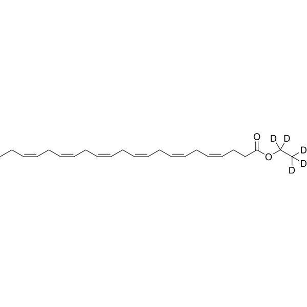
NIR-Red Dead Cell-1 Dye 是一种非活细胞的 DNA 结合荧光染料 (Ex/Em=515 nm/531 nm)。NIR-Red Dead Cell-1 Dye 可嵌入双链 DNA 的碱基对,并产生更强的应该。NIR-Red Dead Cell-1 Dye 适用于细胞膜受损的坏死细胞或凋亡晚期细胞,在荧光显微镜或流式细胞术下呈现绿色荧光。NIR-Red Dead Cell-1 Dye 可用于区分活细胞与死细胞,区分细胞膜完整性。NIR-Red Dead Cell-1 Dye 可以附着在 Feraheme (FH) 纳米颗粒 (NP) 的表面,以获得荧光染料功能化的 NP,用于药物递送的研究。
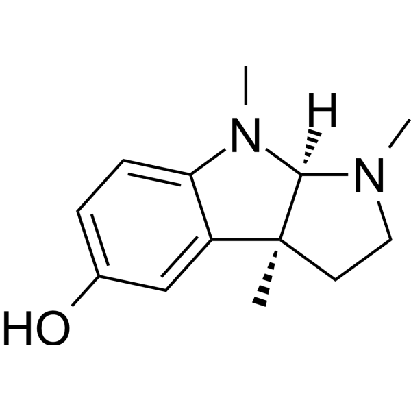
假马齿苋皂素 A
Bacoside A 是一种口服有效、具有血脑屏障通透性的三萜皂苷,能够调节 ATP 酶、AChE、CaMK2A 及 iNOS 的活性。源自 Bacopa monniera。Bacoside A 通过维持离子平衡、清除活性氧、稳定细胞膜以及调控 NF-κB 和凋亡相关蛋白表达,发挥显著的抗氧化、抗炎和抗凋亡作用。Bacoside A 可对抗吗啡诱导的 Na+/K+-ATPase、Ca2+-ATPase 和 Mg2+-ATPase 活性降低,提高线粒体膜电位,降低细胞内活性氧水平。Bacoside A 能特异性结合钙/钙调蛋白依赖性蛋白激酶 IIA 触发内质网钙释放。Bacoside A 对胶质母细胞瘤细胞表现出非凋亡性细胞毒性,同时保护正常神经细胞免受应激损伤。Bacoside A 可用于帕金森病及多形性胶质母细胞瘤的研究。
寡霉素 B
Oligomycin B 是一种抗生素 (antibiotic),可作为 ATP 合酶 (ATP Synthase) 的非选择性抑制剂。Oligomycin B 可升高线粒体膜电位。Oligomycin B 诱导细胞凋亡 (apoptosis) 和坏死。Oligomycin B 会削弱葡萄霜霉菌游动孢子的运动能力并诱导其裂解。Oligomycin B 抑制小麦稻瘟病菌,并抑制小麦瘟病的发生。Oligomycin B 可降低灰葡萄孢菌的菌丝生长与孢子萌发水平,保护拟南芥抵御灰葡萄孢菌的侵害。Oligomycin B 会加重大脑皮质挫伤大鼠的脑细胞毒性水肿,升高颅内压与脑含水量,并加剧其线粒体损伤。Oligomycin B 可用于葡萄霜霉病、创伤性脑损伤、小麦瘟病、灰霉病的相关研究。
NIR-Red Dead Cell-1 Dye 是一种非活细胞的 DNA 结合荧光染料 (Ex/Em=515 nm/531 nm)。NIR-Red Dead Cell-1 Dye 可嵌入双链 DNA 的碱基对,并产生更强的应该。NIR-Red Dead Cell-1 Dye 适用于细胞膜受损的坏死细胞或凋亡晚期细胞,在荧光显微镜或流式细胞术下呈现绿色荧光。NIR-Red Dead Cell-1 Dye 可用于区分活细胞与死细胞,区分细胞膜完整性。NIR-Red Dead Cell-1 Dye 可以附着在 Feraheme (FH) 纳米颗粒 (NP) 的表面,以获得荧光染料功能化的 NP,用于药物递送的研究。
寡霉素 B
Oligomycin B 是一种抗生素 (antibiotic),可作为 ATP 合酶 (ATP Synthase) 的非选择性抑制剂。Oligomycin B 可升高线粒体膜电位。Oligomycin B 诱导细胞凋亡 (apoptosis) 和坏死。Oligomycin B 会削弱葡萄霜霉菌游动孢子的运动能力并诱导其裂解。Oligomycin B 抑制小麦稻瘟病菌,并抑制小麦瘟病的发生。Oligomycin B 可降低灰葡萄孢菌的菌丝生长与孢子萌发水平,保护拟南芥抵御灰葡萄孢菌的侵害。Oligomycin B 会加重大脑皮质挫伤大鼠的脑细胞毒性水肿,升高颅内压与脑含水量,并加剧其线粒体损伤。Oligomycin B 可用于葡萄霜霉病、创伤性脑损伤、小麦瘟病、灰霉病的相关研究。
假马齿苋皂素 A
Bacoside A 是一种口服有效、具有血脑屏障通透性的三萜皂苷,能够调节 ATP 酶、AChE、CaMK2A 及 iNOS 的活性。源自 Bacopa monniera。Bacoside A 通过维持离子平衡、清除活性氧、稳定细胞膜以及调控 NF-κB 和凋亡相关蛋白表达,发挥显著的抗氧化、抗炎和抗凋亡作用。Bacoside A 可对抗吗啡诱导的 Na+/K+-ATPase、Ca2+-ATPase 和 Mg2+-ATPase 活性降低,提高线粒体膜电位,降低细胞内活性氧水平。Bacoside A 能特异性结合钙/钙调蛋白依赖性蛋白激酶 IIA 触发内质网钙释放。Bacoside A 对胶质母细胞瘤细胞表现出非凋亡性细胞毒性,同时保护正常神经细胞免受应激损伤。Bacoside A 可用于帕金森病及多形性胶质母细胞瘤的研究。
Western blot analysis of extracts from THP-1(lane 2(20μg), Jurkat (lane 3(20μg) and NIH3T3(lane 4(20μg) using FOXO1A (HY-P80132) Rabbit mAb. Proteins were transferred
to a PVDF membrane and blocked with 5% non-fat milk in TBST for 2 hour at room temperature. The primary antibody (1/1000) and Loading control antibody (Beta Actin, HY-P80438, 1/10000) was
used in 5% non-fat milk in TBST at 4°C overnight. Goat Anti-Mouse/Rabbit IgG-HRP Secondary Antibody (1/10000) was used for 1 hour at room temperature.
Western blot analysis of extracts from THP-1(lane 2(20μg), Jurkat (lane 3(20μg) and NIH3T3(lane 4(20μg) using FOXO1A (HY-P80132) Rabbit mAb. Proteins were transferred
to a PVDF membrane and blocked with 5% non-fat milk in TBST for 2 hour at room temperature. The primary antibody (1/1000) and Loading control antibody (Beta Actin, HY-P80438, 1/10000) was
MedchemExpress Validation 03
Western blot analysis of extracts from THP-1(lane 2(20μg), Jurkat (lane 3(20μg) and NIH3T3(lane 4(20μg) using FOXO1A (HY-P80132) Rabbit mAb. Proteins were transferred
MedchemExpress Validation 04
Western blot analysis of extracts from THP-1(lane 2(20μg), Jurkat (lane 3(20μg) and NIH3T3(lane 4(20μg) using FOXO1A (HY-P80132) Rabbit mAb. Proteins were transferred
to a PVDF membrane and blocked with 5% non-fat milk in TBST for 2 hour at room temperature. The primary antibody (1/1000) and Loading control antibody (Beta Actin, HY-P80438, 1/10000) was
used in 5% non-fat milk in TBST at 4°C overnight. Goat Anti-Mouse/Rabbit IgG-HRP Secondary Antibody (1/10000) was used for 1 hour at room temperature.
MedchemExpress Validation
Western blot analysis of extracts from THP-1(lane 2(20μg), Jurkat (lane 3(20μg) and NIH3T3(lane 4(20μg) using FOXO1A (HY-P80132) Rabbit mAb. Proteins were transferred
to a PVDF membrane and blocked with 5% non-fat milk in TBST for 2 hour at room temperature. The primary antibody (1/1000) and Loading control antibody (Beta Actin, HY-P80438, 1/10000) was
used in 5% non-fat milk in TBST at 4°C overnight. Goat Anti-Mouse/Rabbit IgG-HRP Secondary Antibody (1/10000) was used for 1 hour at room temperature.
MedchemExpress Validation
Western blot analysis of extracts from THP-1(lane 2(20μg), Jurkat (lane 3(20μg) and NIH3T3(lane 4(20μg) using FOXO1A (HY-P80132) Rabbit mAb. Proteins were transferred
to a PVDF membrane and blocked with 5% non-fat milk in TBST for 2 hour at room temperature. The primary antibody (1/1000) and Loading control antibody (Beta Actin, HY-P80438, 1/10000) was
used in 5% non-fat milk in TBST at 4°C overnight. Goat Anti-Mouse/Rabbit IgG-HRP Secondary Antibody (1/10000) was used for 1 hour at room temperature.